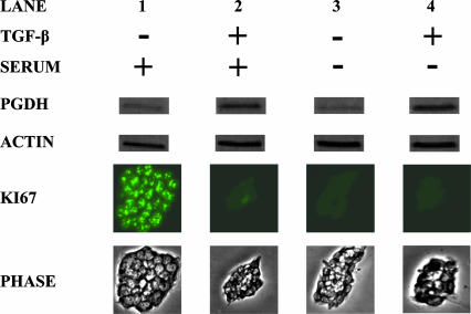
Fig. 5.
15-PGDH induction by TGF-β but not by cell growth arrest. Shown is Western blot assay of 15-PGDH protein expression in FET cells treated with TGF-β (10 ng/ml) (TGF-β +) versus control (TGF-β –) and maintained in basal medium plus 8% serum (serum +) or minus the addition of (serum –). Proliferative status of the cells is determined by immunofluorescent assay for Ki67, and colony size was visualized by phase microscopy. Assays were performed on day 5 of cell culture.

