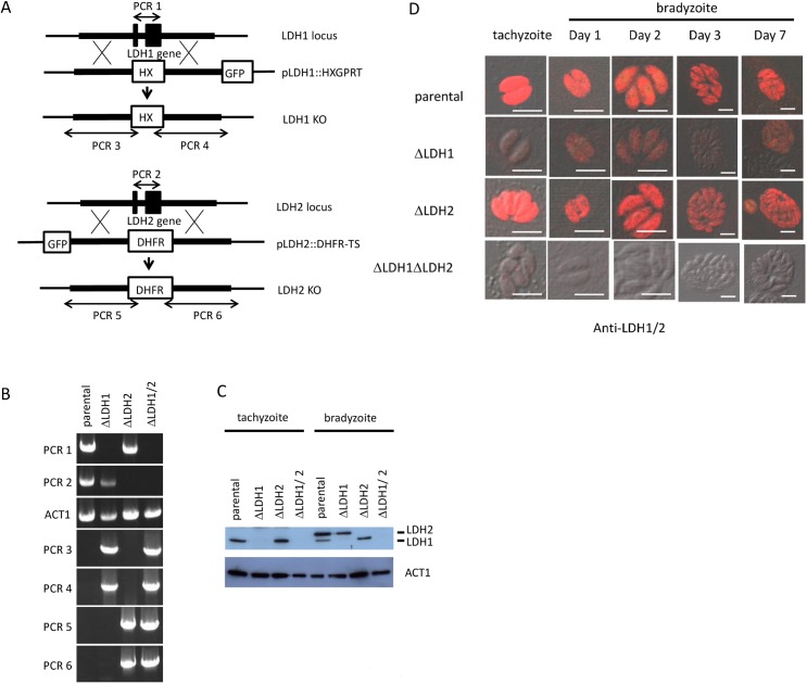
Fig 1. Construction of knockout strains.
(A) Schematic diagram of LDH gene disruption strategy by double crossover homologous recombination in type II PruΔku80Δhxgprt using a plasmid, pΔLDH containing the 5’ and 3’ flanking region. HXGPRT and DHFR-TS selectable markers were used for selection. (B) PCR analysis for validation of gene knockouts. Signal of coding region of LDH1 (PCR1) is absent in Δldh1 and Δldh1Δldh2 strains. Signal of coding region of LDH2 (PCR2) is absent in Δldh2 and Δldh1Δldh2 strains. The actin coding region (ACT1) was used as a PCR control. Successful homologous recombination is indicated by the presence of signal of either LDH1 (PCR3 and 4) in Δldh1 and Δldh1/2 or LDH2 (PCR5 and 6) in Δldh2 and Δldh1/2 strains. (C) Western blot analysis of parental and knockout parasite lysates of tachyzoite and bradyzoite stage. Anti-LDH1/2 antibody which recognizes both LDH1 and 2 proteins was used. LDH1 signal is absent in Δldh1 and Δldh1/2 strains and LDH2 signal is absent in Δldh2 and Δldh1/2 strains. Although the predicted molecular weight of LDH1 and 2 is nearly identical (35.5 and 35.3 kDa, respectively), western blot antibody reactive bands of LDH2 migrated at an apparent higher molecular weight than LDH1. (D) Immunofluorescence staining of parental and knockout parasite lysates of tachyzoite and bradyzoite stage. Anti-LDH1/2 antibody which recognize both LDH1 and 2 proteins are used. In the LDH1 disruption, weak signal gradually appeared after bradyzoite induction. No signal was detected in the candidate LDH1/LDH2 double knockout parasites. White bar in photos indicates 10μm.

