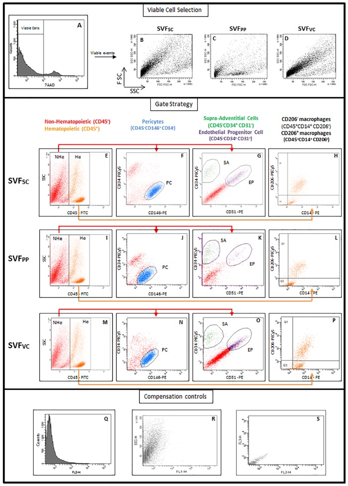
Fig 1. Perivascular and hematopoietic populations classified for analytical flow cytometry.
SVF of subcutaneous, preperitoneal and visceral adipose tissues from morbidly obese patients was analyzed by flow cytometry. First, viable cells were identified by 7AAD exclusion (A). Second, viable cells were distributed in a Forward versus Side Scatter plot (B, C, D) and further analyzed according to CD45 expression (E, I, M). The gates were set on non-hematopoietic (CD45neg—NHe) and hematopoietic (CD45pos—He) cells. Third, CD45neg cells (red) were analyzed for CD34 and CD146 expression (F, J, N) or CD34 and CD31 expression (G, K, O). A blue gate was set on the CD146posCD34neg cells to identify the pericytes. A subset of supra-adventitial cells (SA), which were CD34posCD31neg, was identified (green gate) whereas the endothelial progenitor cells (EP) were identified as CD34posCD31pos (purple gate). Among the He cells, two monocyte-macrophage populations were identified (H, L, P): adipose tissue resident macrophages were identified as CD14posCD206pos cells (upper right quadrant) and a population of CD14posCD206neg cells (lower right quadrant). (Q-S) Compensation controls of fluorescence detection. SVF: stromal vascular fraction; SC: Subcutaneous; PP: preperitoneal; VC: visceral; He: hematopoietic; NHe: non-hematopoietic; PC: pericytes; SA: supra-adventitial cells; EP: endothelial progenitor cell.

