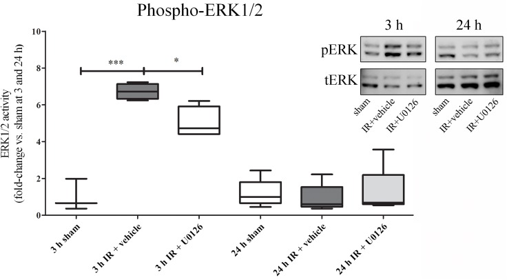
Fig 2. Western blot analysis of heart homogenates that contained myocardium and the left anterior descending artery (LAD).
A. There was a significant increase in ERK1/2 phosphorylation as normalized to the total ERK1/2 at 3 h of reperfusion, and this was significantly attenuated by treatment with U0126. After 24 h, phospho-ERK levels had returned to baseline and no changes were seen between the groups. Western blot results are shown as dot plots, where each dot represent an individual rat (n = 4–5). Bars and whiskers indicate mean values ± SEM. *P < 0.05, ***P < 0.001, one-way ANOVA with Bonferroni’s multiple comparison test.

