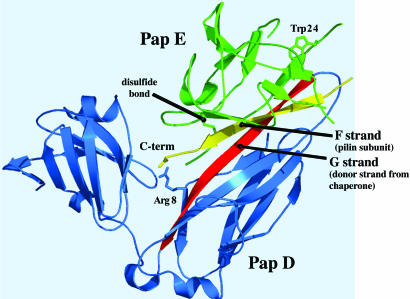
Fig. 1.
X-ray structure of PapD(6-His)-PapENTD (20). PapD is in blue, and the subunit PapENTD is in green, with the disulfide and tryptophans highlighted. The G strand of the chaperone that occupies the groove of the subunit is shown in red, and the C-terminal strand of the subunit that binds to Arg-8 in the chaperone cleft is shown in yellow. The figure was generated with pymol (Delano Scientific, San Carlos, CA).

