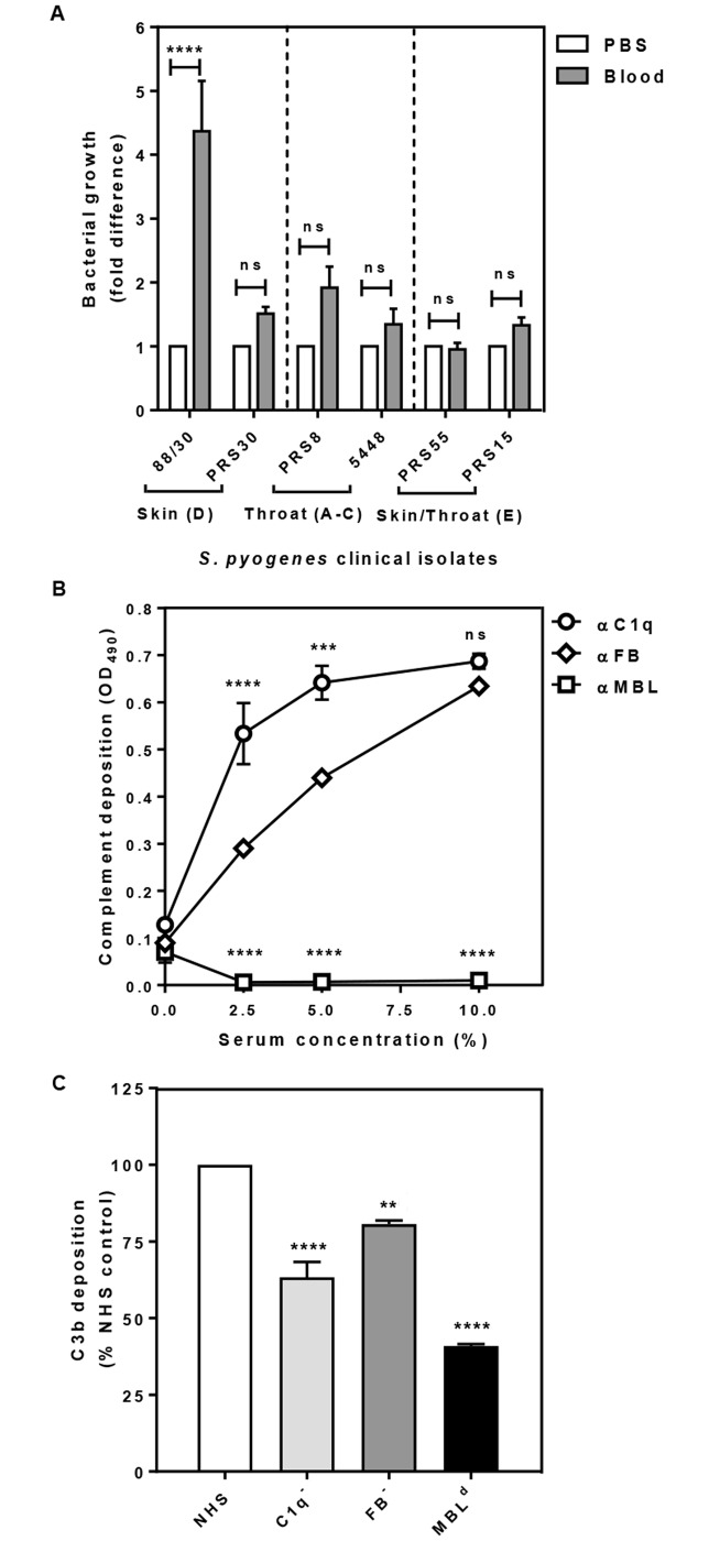
Fig 1. GAS clinical isolates are naturally resistant to blood killing (A) and deposition of C1q, MBL, FB (B) and C3b, indicative of opsonisation (C) on the cell surface of GAS 88/30.
Skin strains 88/30, PRS30 (emm cluster D), throat strains PRS8, 5448 (emm cluster A-C) and skin/throat strains PRS55, PRS15 (emm cluster E) were harvested from mid-log growth phase culture (OD600 0.35). Suspension of GAS in PBS (1 ×10³ cfu/ml) were added into fresh blood pre-treated with GVB2+ buffer. After 3 h incubation samples were plated in duplicate on CBAC agar plates and bacteria were enumerated as cfu/ml. GAS cell in PBS without blood at time 0 (T0) was plated simultaneously and the numbers of bacteria grown served as the baseline for normalisation and for calculating the fold difference of bacteria numbers grown from the experimental samples (A). Maxisorp 96-well plates coated with GAS cells were incubated with increasing concentrations of NHS (B) or with 5% NHS or sera depleted of either C1q (C1-) or FB (FB-) and serum naturally deficient in MBL (MBLd) (C). Complement deposition was detected by ELISA using primary human specific antibodies, followed by HRP-conjugated secondary antibodies, and fluorescence was detected at 490 nm (B, C). Data represent the means ± SEM from three independent experiments. The statistical significance of differences between samples was estimated using two way ANOVA with Tukey’s multiple comparison test (A, B) and one way ANOVA with Dunnett’s multiple comparison tests (C). **, p<0.01; ***, p<0.001; ****, p<0.0001, ns, not significant.

