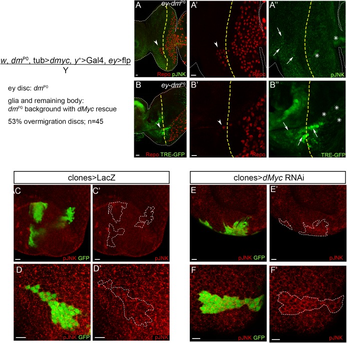
Fig 4. Clonal inhibition of dMyc induces JNK pathway activation.
(A and B) Glia overmigration and JNK activation in eye discs mutant for Myc (ey-dmP0) in a phenotypically wild-type animal. (A) pJNK expression (green) in male dMyc mutant eye discs (ey-dmP0). A’ (glia) and A” (pJNK) show a magnification from A. (B) TRE-GFP expression (green) in male dMyc mutant eye discs (ey-dmP0). B’ (glia) and B” (TRE-GFP) show a magnification from B. Arrowheads point towards glia overmigration (beyond the MF). Arrows indicate eye disc areas with high JNK pathway activation and asterisk represent JNK activation in glia. Glial cells stained with repo are shown in red. A yellow dashed line represents the MF. Scale bars correspond to 10 μm. (C–F) Control (C and D; LacZ) or dMycRNAi (E and F) clones were induced in the Drosophila early eye disc 48 ± 24 hours after egg laying (AEL). Images show representative clones in the epithelial layers of the disc proper (C and E) and peripodial epithelium (D and F), marked positively by the presence of GFP. pJNK is shown in red. A dashed white line surrounds the clone positive region. Scale bars correspond to 10 μm.

