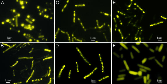
Fig. 1.
Subcellular distribution of FP under different growth conditions. Fluorescence micrographs of BL21-DE3-ΔEI cells harboring pRSETB-Y-EI plasmid grown on agar plates containing LB medium (A) or Neidhardt medium supplemented with 20 mM d-mannitol (B), 20 mM d-ribose (C), or 40 mM dl-lactate (D). (E and F) Cells in liquid culture. Shown are 20 mM d-ribose in Neidhardt medium (E) and control cells harboring pRSETB-YFP (no enzyme I) in LB broth in the presence of 0.5 mM IPTG (F). See Results.

