Figure 3.
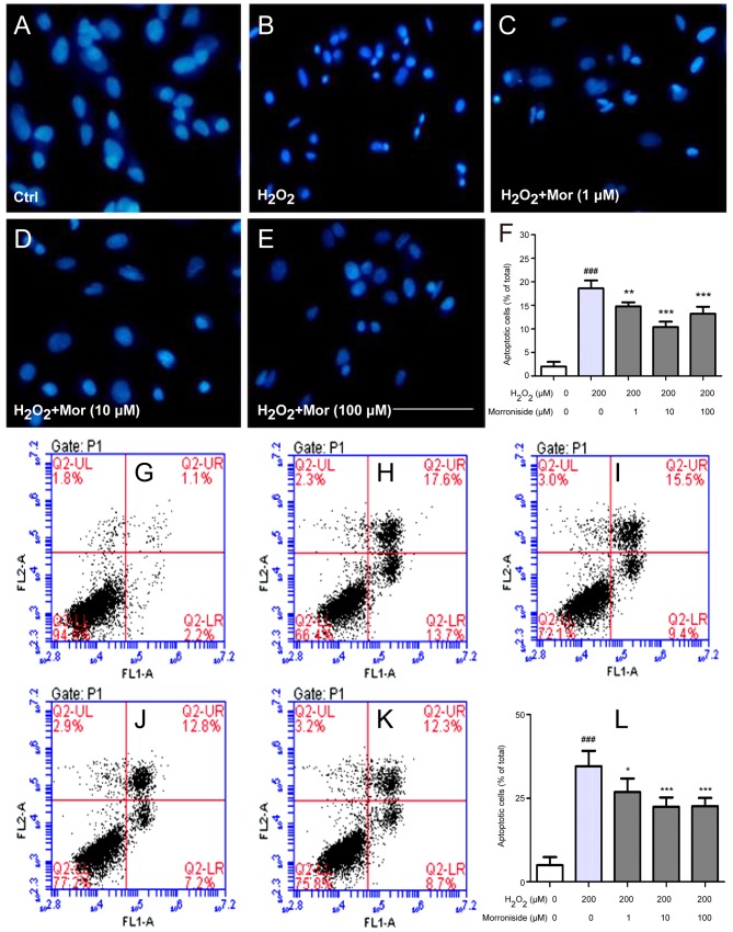
Morroniside blocks H2O2-induced apoptosis of SK-N-SH cells. Apoptotic nuclei were visualized by Hoechst 33342 staining. (A) Untreated cells with normal nuclei. (B) Cells exposed to 200 µM H2O2 with condensed nuclei. (C–E) Cells pretreated with morroniside at concentrations of (C) 1 µM, (D) 10 µM and (E) 100 µM for 24 h showed reduced sensitivity to the effects of H2O2, as evidenced by fewer cells with condensed nuclei. Scale bar, 20 µM. (G–K) Annexin V/propidium iodide (PI) double-staining was used to confirm apoptosis in SK-N-SH cells. Few cells positive for Annexin V/PI staining were detected in the control group (G), while H2O2 (200 µM) treatment increased the percentage of Annexin V+/PI+ cells (H). Preincubation with (I) 1 µM, (J) 10 µM and (K) 100 µM morroniside for 24 h inhibited H2O2-induced apoptosis. Quantitative analyses are shown in panels (F and L). Data represent mean ± SD of three independent experiments. ###P<0.001 vs. control group; *P<0.01, **P<0.01 and ***P<0.001 vs. injury group.

