Figure 2.
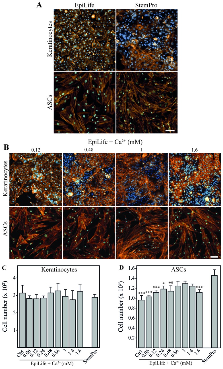
The effect of calcium on morphology and cell number of keratinocytes and adipose-derived stem cells (ASCs). Keratinocytes and ASCs were cultured until 80% confluent, followed by 24 h of incubation in EpiLife, EpiLife supplemented with calcium, or StemPro. (A) Representative images of keratinocytes and ASCs showing morphology and cell density after incubation in EpiLife or StemPro. To assess cell morphology and cell number, cytoskeleton were visualized by phalloidin-Bodipy 558/568 staining (orange) and nuclei were counterstained with Hoechst 33342 (blue). The scale bars denote 100 µm. (B) Representative images of keratinocytes and ASCs showing morphology and cell density after incubation in EpiLife supplemented with increasing concentrations of calcium. Assessment of morphology was performed as described for (A). (C) Number of keratinocytes after incubation in EpiLife (Ctrl), EpiLife supplemented with increasing concentrations of calcium, or StemPro. Values are represented as the means and SEM; no statistically significant differences were found (n=6). (D) Number of ASCs after incubation in EpiLife, EpiLife supplemented with increasing concentrations of calcium, or StemPro. Values are represented as mean and SEM (n=6). The data from the EpiLife-based media were compared to those from StemPro using a one-way ANOVA followed by a multiple comparisons vs. control, *p<0.05, **p<0.01 and ***p<0.001.

