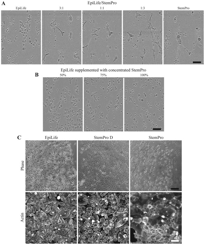
Figure 3.
Effect on keratinocyte morphology of 24 h culture in combinations of EpiLife and StemPro. (A) Keratinocytes were cultured in either EpiLife, StemPro, or combinations of EpiLife and StemPro in the ratios of 3:1, 1:1 and 1:3, after which the morphology was assessed by phase contrast microscopy. The scale bars denote 150 µm. (B) The protein fraction from conditioned StemPro was concentrated on spin columns, reconstituted in EpiLife at 50, 75 and 100% of the original concentration, and added to keratinocytes. The morphology was assessed by phase contrast microscopy as above. (C) Keratinocytes were cultured in either EpiLife, StemPro dialyzed against EpiLife (StemPro D), or StemPro. The morphology was assessed by phase contrast microscopy. The scale bars denote 200 µm. To assess the cytoskeleton, actin fibers were visualized by fluorescence microscopy using a phalloidin-Bodipy 558/568 staining. The scale bars denote 100 µm.

