Figure 2.
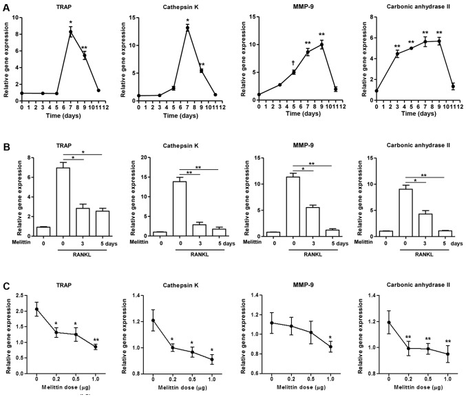
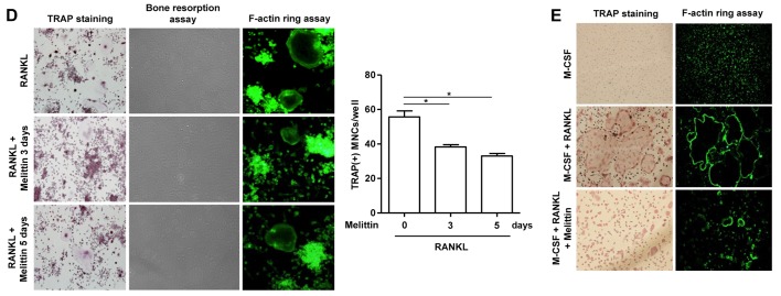
Melittin influencea osteoclast formation in time- and dose-dependent manner. (A) Osteoclastogenic genes, such as TRAP, cathepsin K, matrix metalloproteinase-9 (MMP-9) and carbonic anhydrase II, in the course of osteoclastogenesis in RAW 264.7 cells incubated with receptor activator of nuclear factor-κB ligand (RANKL) (50 ng/ml) for 0, 3, 5, 7, 9 and 11 days were measured (*p<0.001, **p<0.01 and †p<0.05 compared to non-treated cells). (B) Melittin (1 µg/ml) was added to the RAW 264.7 cells on days 3 and 5 following stimulation with RANKL. Inhibitory effect of melittin (1 µg/ml) treatment for 0, 3 and 5 days on osteoclastogenic genes was assessed (*p<0.01 and **p<0.001 compared to RANKL-stimulated cells without melittin). (C) RAW 264.7 cells treated with RANKL (50 ng/ml) were cultured at different dosages of melittin (0.2 to 1.0 µg/ml) (*p<0.05 and **p<0.01 compared to non-treated cells). (D) Melittin was added to the RAW 264.7 cells on days 3 and/or 5 following stimulation with RANKL. The in vitro morphological and functional analyses for osteoclastogenesis were performed using TRAP staining, pit formation, and F-actin ring staining assays in RAW 264.7 cells treated with RANKL with or without melittin (*p<0.01 compared to non-treated cells) (E) The assessment of osteoclast-like cells differentiated from bone marrow-derived macrophages (BMMs) was done using TRAP staining and F-actin ring staining assays. Data are representative of 3 independent experiments.


