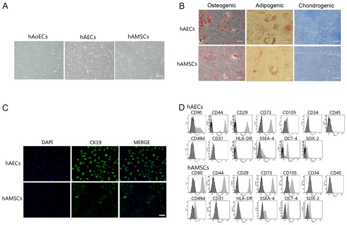
Figure 1.
Characterization of human amniotic epithelial and mesenchymal stem cells. (A) Representative images of human aortic endothelial cells (hAoECs) (passage 4) and cultured cells obtained from human amniotic epithelial (passage 2) and mesenchymal (passage 4) stem cells. Scale bar, 200 μm. (B) Differentiation of amniotic cells into osteocytes (scale bars, 200 μm), adipocytes (scale bars, 20 μm) and chondrocytes (scale bars, 200 μm). Cells cultured under osteogenic, adipogenic or chondrogenic culture conditions were stained for calcium deposits with alizarin red staining, lipid droplets with Oil Red O staining or proteoglycans with Alcian blue staining, respectively. (C) Immunofluorescence was conducted to evaluate cytokeratin 19 expression in human amniotic epithelial cells (hAECs) and human amniotic mesenchymal stem cells (hAMSCs). Scale bar, 100 μm. (D) FACS analysis of cell markers of hAECs and hAMSCs (light gray bars) compared with isotype-matched antibodies (dark gray bars).

