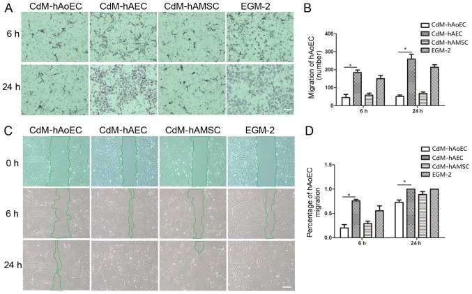
Figure 3.
Effect of conditioned medium (CdM) on the migration of human aortic endothelial cells (hAoECs). (A) Images of hematoxylin and eosin-stained membranes in the Transwell hAoEC migration assay after a 6- and 24-h coculture with CdM. (B) Rate of hAoEC movement. Results indicate that hAoEC migration into the bottom chamber was accelerated in the presence of CdM-hAEC. Date represent the mean ± SD of there independent experiments. *P<0.05 (C) Migration of hAoECs into the scratch wound after 0, 6 and 24 h of culture with CdM in the scratch wound assay. (D) Rate of hAoEC movement after 6 and 24 h. The rate of movement was signifiantly greater for hAoECs cultured with CdM-hAEC compared with the other groups. Scale bar, 200 μm. CdM-hAoEC (control), CdM from hAoECs; CdM-hAEC, CdM from human amniotic epithelial cells; CdM-hAMSC, CdM from human amniotic mesenchymal stem cells.

