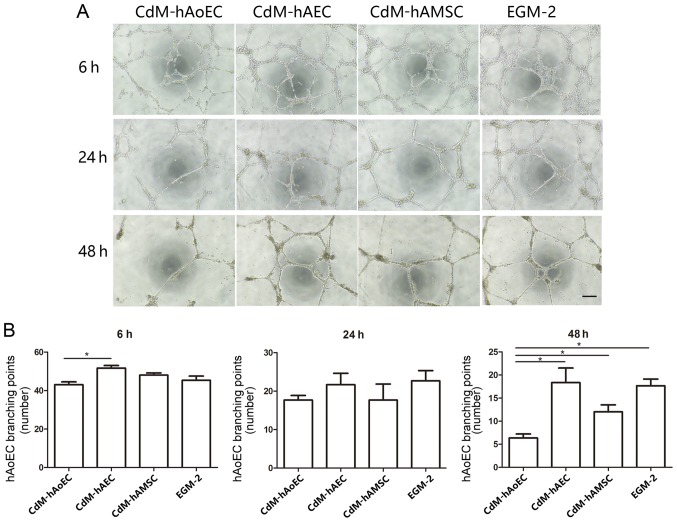
Figure 4.
Matrigel tube formation analysis in vitro. (A) Representative images of Matrigel tube formation using conditioned medium (CdM) at 6, 24 and 48 h. CdM-hAoEC was used as a negative control. Scale bar, 200 μm. (B) Representation of the branching point number of hAoECs. The number was significantly higher in the CdM-hAEC and CdM-hAMSC group. *P<0.05. CdM-hAoEC, CdM from human aortic endothelial cells; CdM-hAEC, CdM from human amniotic epithelial cells; CdM-hAMSC, CdM from human amniotic mesenchymal stem cells.

