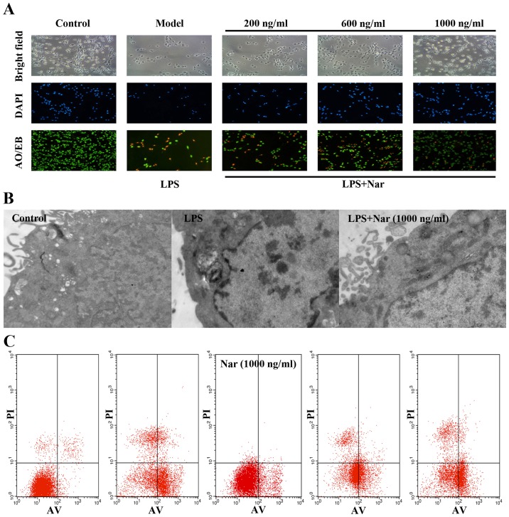
Figure 2.
(A) Morphological and fluorescence images of PC12 cells stained with acridine orange and ethidium bromide AO/EB and DAPI (×100 magnification). (B) Protective effect of the naringin (Nar) on the ultra-structure of PC12 cells (×40,000 magnification). (C) Detection of apoptosis by flow cytometry. Data are presented as the means ± SD (n=5 treatment groups). *p<0.05 and **p<0.01 compared with model group.

