. 2017 Mar 21;6:e21130. doi: 10.7554/eLife.21130

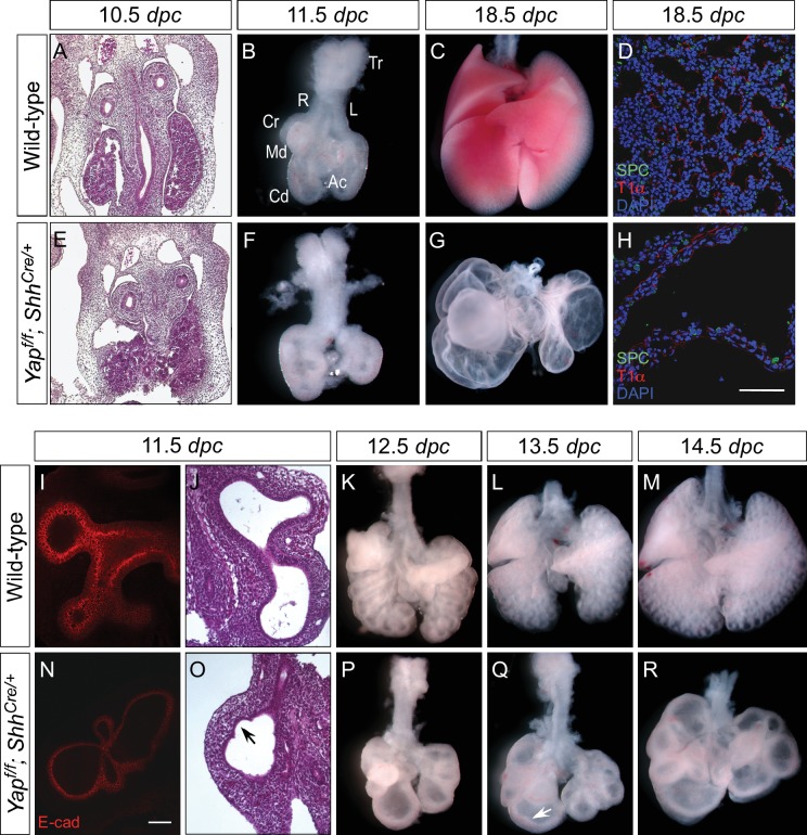
Figure 2. Loss of epithelial Yap leads to lung cysts.

(A,E) Hematoxylin and eosin-stained sections of wild-type and Yapf/f; ShhCre/+ embryos at 10.5 dpc. No apparent difference in the morphology of lung buds was observed between wild-type and Yap mutants. (B,C,F,G) Ventral view of dissected lungs from wild-type and Yapf/f; ShhCre/+ mice at 11.5 and 18.5 dpc. Five buds were produced in both control and Yap-deficient lungs at 11.5 dpc; defective lung branching along the entire lung epithelium could already be detected at this stage in Yap mutants. As lung development proceeded, failure to execute a stereotyped program of branching in the absence of Yap resulted in lungs consisting only of multiple cysts at 18.5 dpc. R, right; L, left; Tr, trachea; Cr, cranial; Md. middle; Cd, caudal; Ac, accessory. (D,H) Immunostaining of lung sections collected from wild-type and Yapf/f; ShhCre/+ mice at 18.5 dpc. Cell types in the proximal airway of Yapf/f; ShhCre/+ mice failed to be specified. For instance, expression of markers for Clara [club] cells (CC10+), ciliated cells (acetylated-tubulin [Ac-tub]+) and pulmonary neuroendocrine cells (CGRP+) were barely detectable (not shown). Reduction in the expression of distal lung cell markers, such as SPC (type II cells) and T1α (type I cells), in the cysts of Yapf/f; ShhCre/+ lungs was also noted. (I,N) Whole-mount immunostaining of wild-type and Yapf/f; ShhCre/+ lungs at 11.5 dpc by two-photon microscopy. Lung epithelium was identified by E-cadherin (E-cad). (J,O) Hematoxylin and eosin-stained sections of wild-type and Yapf/f; ShhCre/+ embryos at 11.5 dpc. The arrow points to ‘evagination’ of epithelial cells in Yap-deficient lung buds. (K–M, P–Q) Ventral view of dissected lungs from wild-type and Yapf/f; ShhCre/+ embryos at the developmental stages indicated. Epithelial ‘evagination’ (arrow in Q) could be seen in Yap-deficient lung buds but they failed to produce new buds subsequently. All views are ventral. Scale bar = 200 μm for C,G; 50 μm for D,H; 100 μm for I,N.

DOI: http://dx.doi.org/10.7554/eLife.21130.006

Figure 2.

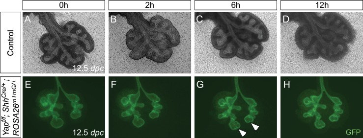
Figure 2—figure supplement 1. Aborted branching in the absence of epithelial Yap in the lung.

Figure 2—figure supplement 1.

(A–H) Time-lapse microscopy of dissected of lungs from control and Yapf/f; ShhCre/+; ROSA26mTmG/+ mice at 12.5 days post coitus (dpc). Lungs were grown on nuclepore membranes for live imaging. At this stage, limited lung branching in Yap-deficient mutants would soon stop. While ‘evaginations’ (arrowheads in G,H) from existing lung buds were noted in the absence of YAP, no new lung buds were generated. Branching occurred in control lungs, but the rate of lung development was slowed down in ex vivo lung explants.
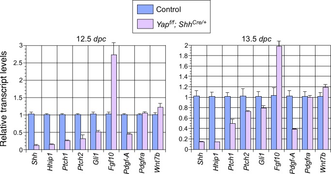
Figure 2—figure supplement 2. Changes in major signaling pathways in the absence of YAP.

Figure 2—figure supplement 2.

qPCR analysis of lungs from control and Yapf/f; ShhCre/+ lungs at 12.5 and 13.5 days post coitus (dpc). The expression levels of Shh were reduced while Fgf10 was upregulated in Yap-deficient lungs. This is consistent with a negative regulatory relationship between Shh and Fgf10. How YAP influences the expression of Shh and Fgf10 and interacts with major signaling pathways requires further investigation. Note that the use of ShhCre has contributed to the reduction in Shh expression levels although it is unlikely to be solely responsible for the low levels of Shh in the absence of Yap.
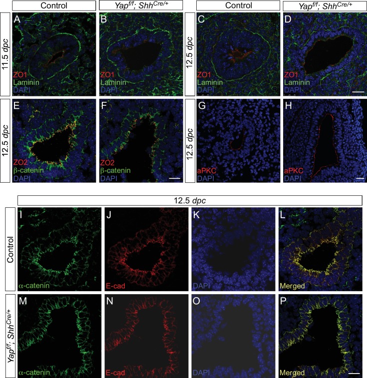
Figure 2—figure supplement 3. Cell junctions and cell polarity are not disrupted due to the loss of epithelial Yap in the lung.

Figure 2—figure supplement 3.

(A–P) Immunostaining of lung sections from control and Yapf/f; ShhCre/+ lungs at 11.5 and 12.5 days post coitus (dpc). No apparent difference in the distribution of apical (aPKC), subapical (ZO1 and ZO2) and basal (Laminin) markers was found between control and Yap mutant lungs. Moreover, the distribution of markers for tight junctions (e.g. ZO1 and ZO2) and adherens junctions (e.g. E-cadherin, β-catenin and α-catenin) was similar between control and Yap-deficient lungs. This suggests that cell junction and cell polarity remain intact in the absence of Yap. Scale bar = 25 μm for A–D, G–H; 100 μm for E–F, I–P.