. 2017 Mar 21;6:e21130. doi: 10.7554/eLife.21130

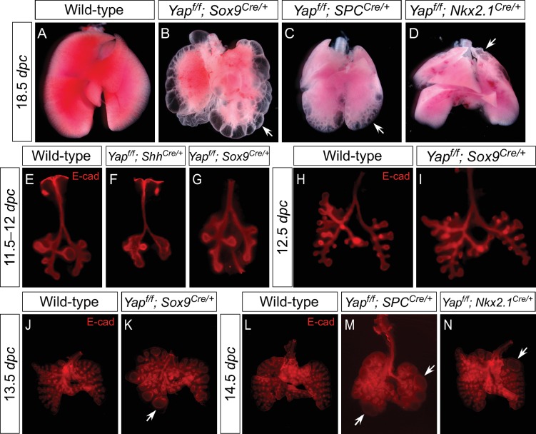
Figure 3. Regional loss of epithelial Yap leads to localized lung cysts.

(A–D) Ventral view of dissected lungs from wild-type, Yapf/f; Sox9Cre/+, Yapf/f; spcCre/+ and Yapf/f; Nkx2.1Cre/+ mice at 18.5 dpc. Lung cysts in Yapf/f; Sox9Cre/+ and Yapf/f; spcCre/+ mice were largely confined to the distal airway (arrows in B,C), while lung cysts were found in the upper lobes (arrow in D) of Yapf/f; Nkx2.1Cre/+ mice. The location of lung cyst formation is correlated with the sites of strong Cre activity and Yap removal. This suggests that loss of Yap at a given region leads to localized lung cysts. (E–N) Whole-mount immunostaining of dissected lungs from wild-type, Yapf/f; ShhCre/+, Yapf/f; Sox9Cre/+, Yapf/f; spcCre/+ and Yapf/f; Nkx2.1Cre/+ mice at the stages indicated. Lung epithelium was visualized by E-cadherin (E-cad). While defective branching was apparent in Yapf/f; ShhCre/+ lungs at 11.5 dpc, branching defects and cyst formation in Yapf/f; Sox9Cre/+ lungs did not appear until 13.5 dpc. Cyst formation in Yapf/f; Sox9Cre/+ lungs was confined to the distal airways (arrow in K). Similarly, cyst formation in Yapf/f; spcCre/+ lungs was detected primarily in the distal airways (arrows in M). By contrast, cyst formation was found in the upper lobes (arrow in N) of Yapf/f; Nkx2.1Cre/+ lungs. This suggests that loss of Yap at a given region leads to localized lung cysts. All views are ventral.

DOI: http://dx.doi.org/10.7554/eLife.21130.010

Figure 3.

Figure 3—figure supplement 1. Deletion of Yap in the distal lung epithelium by the Sox9Cre mouse line.

Figure 3—figure supplement 1.

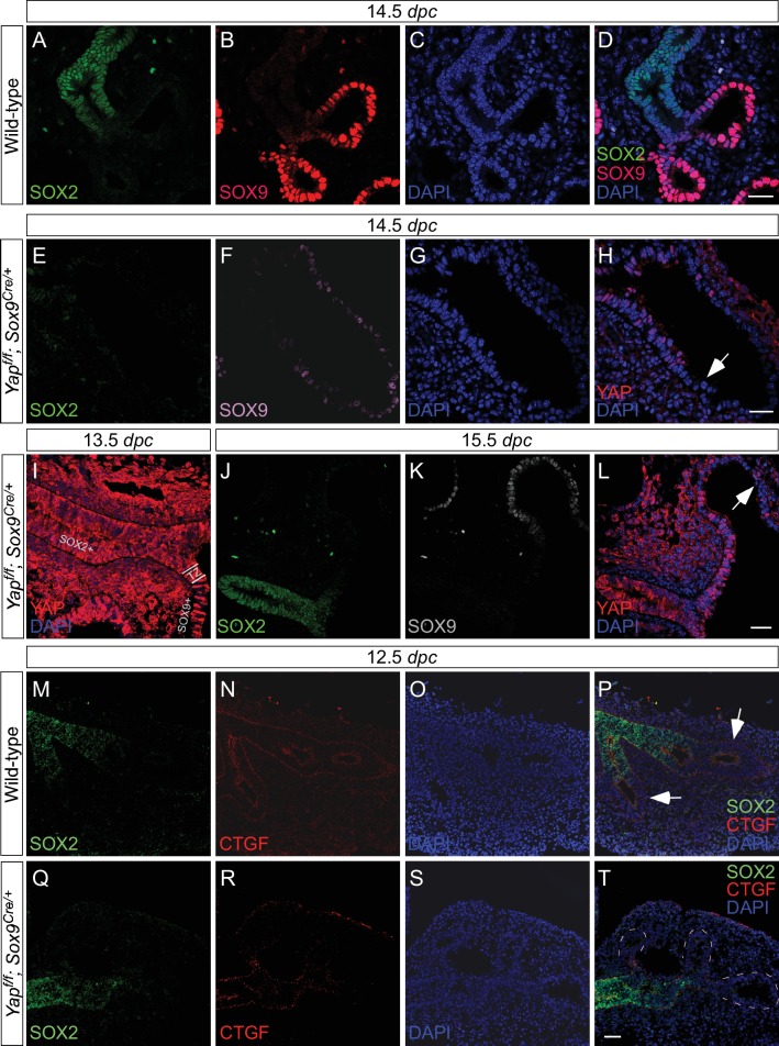
Immunostaining of lung sections collected from wild-type and Yapf/f; Sox9Cre/+ at the stages indicated. (A–D) SOX2 expression marks the proximal airway while the distal airway is distinguished by SOX9 expression. The region where SOX2 and SOX9 expression overlaps is a transition area. (E–L) YAP was lost primarily in distal airways in Yapf/f; Sox9Cre/+ mice, while sporadic loss of YAP can be found in the proximal airway. Loss of YAP was most apparent in the more distal part (arrow in H) of the distal airway, while residual YAP could be found in the more proximal part of the distal airway. (M–T) Removal of YAP in distal airways (dotted lines in T) of Yapf/f; Sox9Cre/+ lungs at 12.5 dpc is associated with loss of CTGF, a direct YAP target. Arrows in P point to CTGF expression in distal airways of wild-type lungs. Together, these results suggest that lung cyst formation in distal airways of Yapf/f; Sox9Cre/+ mice is due to loss of YAP in the distal airway. Scale bar = 25 μm for A–D, E–H; 250 μm for I–L; 100 μm for M–T. dpc, days post coitus.
Figure 3—figure supplement 2. Expression of spcCre is associated with loss of YAP in SOX9+ distal airways.

Figure 3—figure supplement 2.

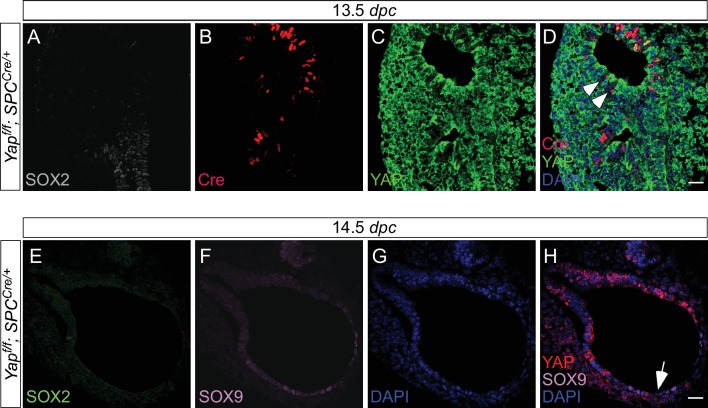
(A–D) Immunostaining of lung sections collected from Yapf/f; spcCre/+ mice at 13.5 days post coitus (dpc). SOX2 expression marks the proximal airway, while the distal airway is distinguished by SOX9 expression (not shown). High levels of spcCre expression were largely confined to the distal airway, where spcCre expression in a given epithelial cell was correlated with loss of YAP immunoreactivity (e.g. arrowheads in D). (E–H) Immunostaining of lung sections collected from Yapf/f; spcCre/+ mice at 14.5 dpc. Only distal airways are shown. YAP was lost mainly in distal airways in Yapf/f; spcCre/+ mice while sporadic loss of YAP was found in the proximal airway. Loss of YAP was most apparent in the more distal part (arrow in H) of the distal airway, while residual YAP could be found in the more proximal part of the distal airway. Together, these results suggest that lung cyst formation in distal airways of Yapf/f; spcCre/+ mice is due to loss of YAP in the distal airway. Scale bar = 25 μm for A–D; E–H.
Figure 3—figure supplement 3. Expression of Nkx2.1Cre is associated with loss of YAP in the upper lobes.

Figure 3—figure supplement 3.

(A–D) Immunostaining of lung sections collected from Yapf/f; Nkx2.1Cre/+ mice at 14.5 days post coitus (dpc). SOX2 expression marks the proximal airway, while the distal airway is distinguished by SOX9 expression (not shown). YAP was lost mainly in the upper lobe in Yapf/f; Nkx2.1Cre/+ lung. Loss of YAP was more apparent in the distal airway, while loss of YAP was sporadic in the proximal airway. Lung cyst formation was primarily observed in the distal airway. The boxed region in (B) indicates areas shown in (C,D). Scale bar = 250 μm for A,B; 250 μm for C,D. Sox2 expression was present in sporadic Yap-deficient cells in the transition zone induced by Sox9Cre, spcCre or Nkx2.1Cre. This suggests that Sox2 expression is not controlled by YAP.
Figure 3—figure supplement 4. Loss of epithelial Yap at restricted areas leads to localized lung cysts.

Figure 3—figure supplement 4.

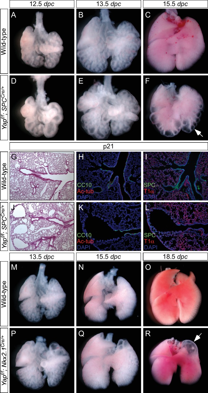
(A–F, M–R) Ventral view of dissected lungs from wild-type, Yapf/f; spcCre/+ and Yapf/f; Nkx2.1Cre/+ mice at various developmental stages as indicated. Lung cysts in Yapf/f; spcCre/+ mice were largely confined to the distal airway (arrow in F), while lung cysts were found in the upper lobes (arrow in R) of Yapf/f; Nkx2.1Cre/+ mice. The location of lung cysts is correlated with the sites of active Cre activity. (G, J) Hematoxylin and eosin-stained sections of lungs from wild-type and occasional adult survivors of Yapf/f; spcCre/+ mice. Multiple cysts in the distal airway were present. (H, I, K, L) Immunstaining of lung sections from control and Yapf/f; spcCre/+ mice. Lung cysts expressed markers of cell types in the distal airway albeit at reduced levels. dpc, days post coitus; p, postnatal.
Figure 3—figure supplement 5. The dosage of Nkx2.1Cre and spcCre affects the severity of lung phenotypes.

Figure 3—figure supplement 5.

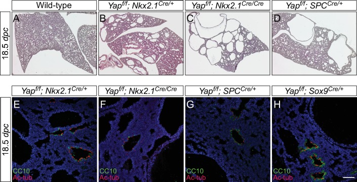
(A–D) Hematoxylin and eosin-stained sections of lungs from wild-type, Yapf/f; Nkx2.1Cre/+ (one copy of Nkx2.1Cre), Yapf/f; Nkx2.1Cre/Cre (two copies of Nkx2.1Cre) and Yapf/f; spcCre/+ (one copy of spcCre) mice at 18.5 dpc. Removal of Yap was more efficient when two copies of Nkx2.1Cre or spcCre were present and lungs phenotypes were more severe. Data not shown for Yapf/f; spcCre/Cre (two copies of spcCre). (E–H) Immunostaining of lung sections collected from Yapf/f; Nkx2.1Cre/+, Yapf/f; Nkx2.1Cre/Cre, Yapf/f; spcCre/+ and Yapf/f; Sox9Cre/+ mice at 18.5 dpc. While more lung cysts were detected in Yapf/f; Nkx2.1Cre/Cre mice than Yapf/f; Nkx2.1Cre/+ mice, cell types in the proximal airway or regions where no cysts were present appeared to be properly specified as indicated by their expression of CC10 (Clara cell marker) and acetylated-tubulin [Ac-tub] (ciliated cell marker). Similarly, cell types in the proximal airway were specified in Yapf/f; spcCre/+ and Yapf/f; Sox9Cre/+ mice. This suggests that lung cyst formation is correlated with Cre activity and is independent of proximal airway specification. Scale bar = 75 μm for E–H. dpc, days post coitus.