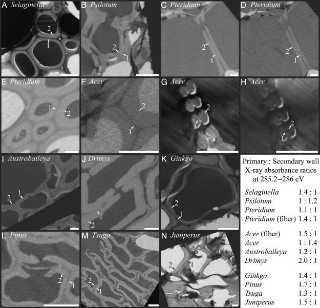
Fig. 1.
X-ray microscopy images of xylem cells with darker shades indicating greater x-ray absorbance and lignin abundance. Images were taken at the 285.2- to 286-eV absorption peak for aromatic carbon except for D and H, which were taken at the 288.5-eV absorption peak for carboxylate carbon most prevalent in pectins. All images are of water-conducting cells except for E and F (sclerenchyma cells). Primary walls (1) and secondary walls (2) are labeled in each image. Nearly black or white areas found in cell lumens are epoxy or holes in the section. (Scale bar, 6 μm.)

