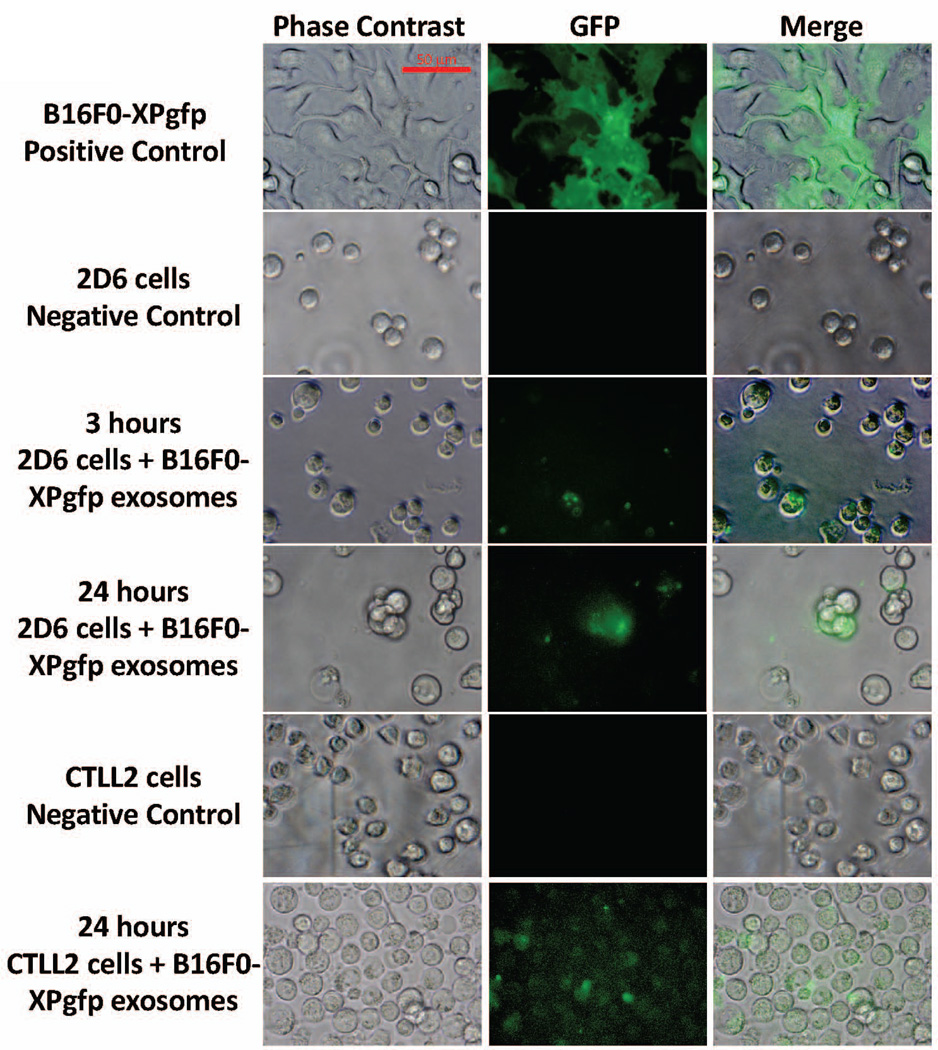
Figure 7. B16F0 exosomes delivered a biological payload to T cells.
B16F0 cells were transfected with a lentivirus-based Xpack-GFP plasmid that targets GFP to exosomes (positive control). Exosomes isolated from B16F0-XPgfp cells were co-cultured with 2D6 (3 and 24 hours) and CTLL2 (24 hours) cells, washed three times with DPBS, and imaged by fluorescence microscopy. Phase contrast and merged images are shown for comparison. 2D6 and CTLL-2 cultured in media alone served as negative controls. Scale bar indicates 50 mum.

