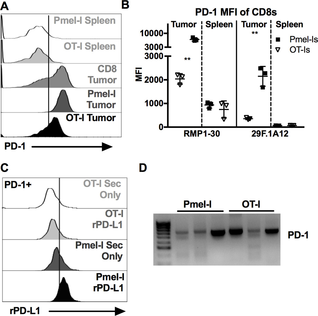
Figure 6. Virus-specific CD8+ TIL express lower amounts of full length PD-1 than tumor-specific CD8+ TIL, altering PD-L1 binding.
500 OT-Is and 5000 Pmel-Is were co-transferred on D-1 and 1 × 10⁵ B16F0s were given on D0. Tumor bearing animals were infected with 2 × 10⁵ pfu of each MCMV-GFP-SL8 and MCMV-GP100 IP on D5. Mice were sacrificed when the tumor reached 100mm2. Shown is (A) representative staining of PD-1 using clone RMP1–30 and (B) average MFI of PD-1 staining using clone RMP1–30 or clone 29F.1A12 for the indicated CD8+ populations recovered from the same tumors (n=3). FACS plots of CD8+ T cells in the spleen include both CD8α IV positive and negative cells while T cells in the tumor tissue include only the CD8α IV negative subset. Significance in (B) was assessed by paired t-tests, p < 0.01 = **. C) Representative binding of recombinant PD-L1 to PD-1+ virus-specific (OT-Is) and TAA-specific (Pmel-Is) recovered from the same tumor (n=4). D) PCR products of PD-1 (Exon 1 through 5) from cDNA derived from sorted virus-specific (OT-Is) and TAA-specific (Pmel-Is) CD8+ TIL (n=3).

