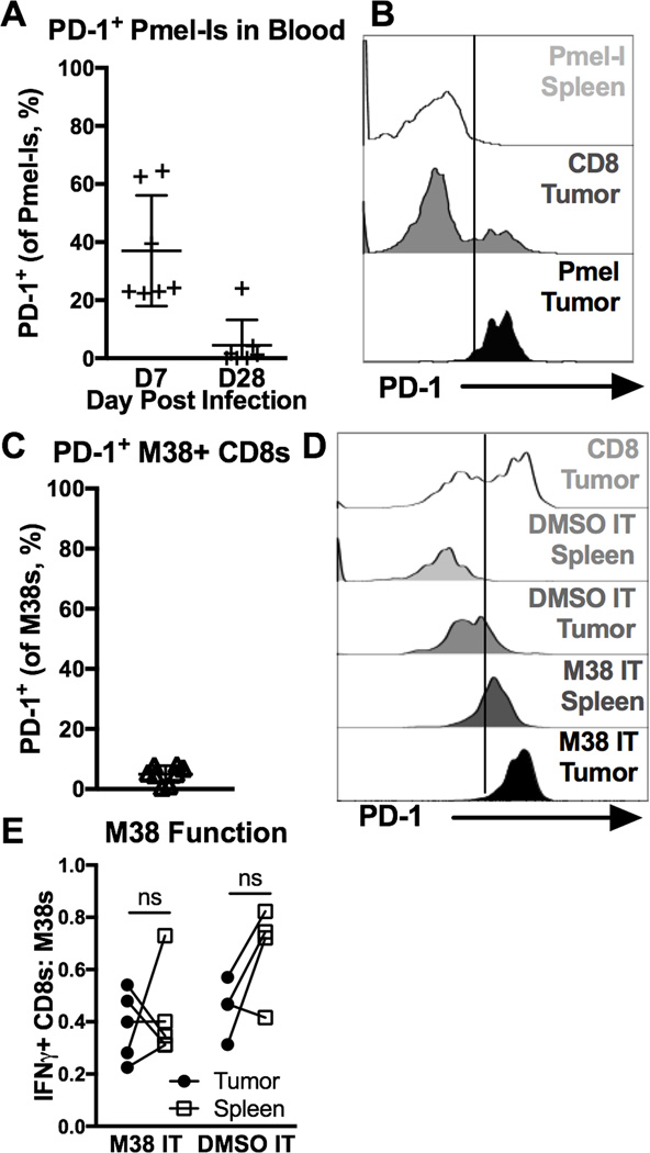
Figure 7. PD-1 is a marker of recent antigen stimulation on TAA-specific and virus-specific CD8+ TIL.
A–B) Animals received 104 Pmel-Is on D-1, were infected with MCMV-gp100 on D0, given B16F0s on D30 or 31, and sacrificed when tumors were ~40mm2 (D14–39, n=6, 2 experiments). Shown is (A) the percent PD-1+ Pmel-Is in the blood D7 and D28 post infection (before tumor implantation), and (B) the MFI of PD-1 expression on total and TAA-specific CD8+ T cells (Pmel-Is) in the tumor and spleen at sacrifice. FACS plots of CD8+ T cells in the spleen include both CD8α IV positive and negative cells while T cells in the tumor tissue include only the CD8α IV negative subset. C-F) Mice infected for 30 days with WT-MCMV were given B16F0s. When tumors were ~20 mm2 they were intratumorally (IT) injected with M38 peptide in PBS (n=5) or DMSO in PBS (n=4), and sacrificed 7 days later (3 experiments). Shown is (C) the percent of PD-1+ M38-specific CD8+ T cells in the blood before tumor implantation, (D) the MFI of PD-1 expression on total and M38-specific CD8+ T cells D7 after IT injections, and (E) the functionality of M38-specific T cells in the tumor and spleen assessed as in Fig. 3B on D7 after IT injections. FACS plots of PD-1 expression in the spleen include both CD8α IV positive and negative cells while T cells in the tumor tissue include only the CD8α IV negative subset. All data is represented as the mean +/− SD and the significance (E) was assessed by paired t-tests, p < 0.05 = *.

