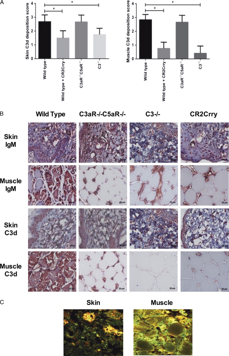
FIGURE 2.

IgM and C3d deposition in vascularized allografts isolated from mice 48 hours after transplantation. A. At 48 hours post-Tx, similar levels of IgM deposition were seen in the skin of VC allografts from all groups, with some decreased deposition in muscle from deficient and inhibited recipients. Mean ± SD, n = 5. *P < 0.05. B. Representative immunohistochemistry images. C. IgM (green) and C3d (red) binding was assessed by immunohistochemistry. Representative immunofluorescence images of WT to WT allografts showing IgM (red) and C3d (green) with colocalization (yellow) n = 3.
