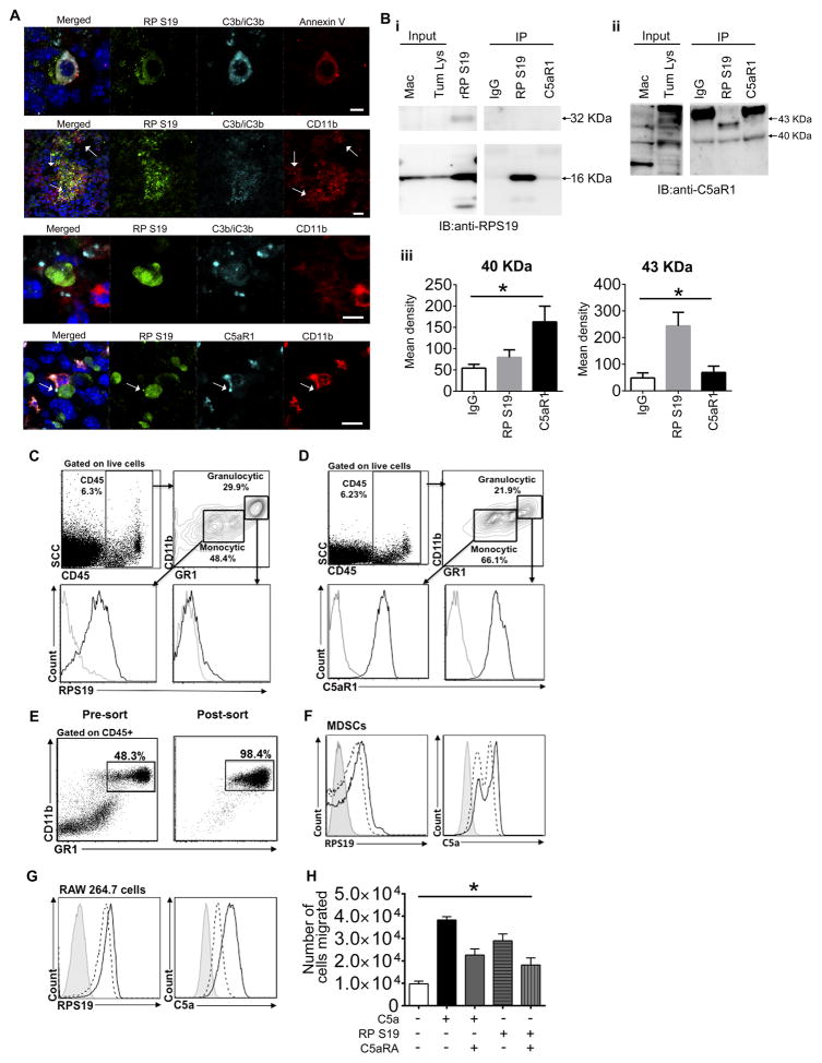
Figure 2. RPS19 binds to C5aR1 expressed on MDSC and macrophages and triggers chemotaxis of macrophages.
(A) RPS19, C3b/iC3b, Anexin V, CD11b, and C5aR1 in murine tumors generated in FVB/N Her2/neu transgenic mouse by injecting NT-5 cells. Arrows: accumulation of CD11b+ cells (upper middle) and co-localization of RPS19 with C5aR1 (lowest panel), scale bars 5 μm except upper middle panel which is 25 μm. (B-i, ii) Immunoprecipitation of RPS19-C5aR1 complex from whole cell lysate (Tum. Lys.) of NT-5 tumors from Her2/neu transgenic mouse. Murine macrophages RAW 264.7 (Mac) were used a positive control for C5aR1 expression and recombinant RPS19 (rRP S19) as control for RPS19. (iii) Quantification of data from ii, *P=0.0364 (right) and *P=0.003 (left) by One-Way-ANOVA. FACS analysis of (C) RPS19 on the surface of monocytic (Gr-1int) vs. granulocytic (Gr-1high) MDSC and (D) C5aR1 expression on monocytic vs. granulocytic MDSC. Black lines: RPS19 and C5aR1 antibodies. Grey lines: isotype controls. (E) Pre- and post-sort FACS of spleen MDSC from NT-5 tumor-bearing mice. Biding of fluorescently labeled-RPS19 or C5a in the absence (continuous black lines) or presence (dashed lines) of C5aR1 antagonist (C5aRA) to (F) sorted MDSC, P<0.0001-RPS19, P=0.0001-C5a by One-Way ANOVA or (G) murine macrophages, P<0.0001-RPS19 and C5a by One-Way ANOVA. (H) Chemotaxis assay; y-axis number of murine macrophages (RAW 264.7) in lower chamber at the end of experiment, *P<0.0001 by One-Way ANOVA. Data are representative of one experiment with n=5 for A, three independent experiments for B–G, and four replicates for H.

