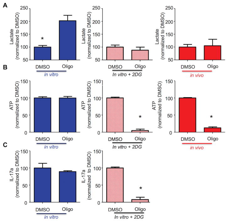
Figure 4. In vivo-differentiated TH17 cells have restricted glycolytic capacity.
(A–C) In vivo-differentiated TH17 (red), in vitro TH17 (blue), and in vitro TH17 treated with 2-deoxyglucose (red stripes) were treated for one hour with DMSO or oligomycin (1μM) and then stimulated with PMA/ionomycin. Lactate concentration in cell-free supernatant from triplicate cultures was evaluated by a colorimetric assay (A), intracellular ATP triplicate cultures by bioluminescence (B), and IL-17 production in cell-free supernatant from triplicate cultures was analyzed by ELISA (C). (A–C) Results are expressed as percentage of DMSO-treated cells. (A–C) Representative of three independent experiments; *p < 0.001 (unpaired Student’s t-test); (error bars, mean ± SEM).

