FIGURE 3.

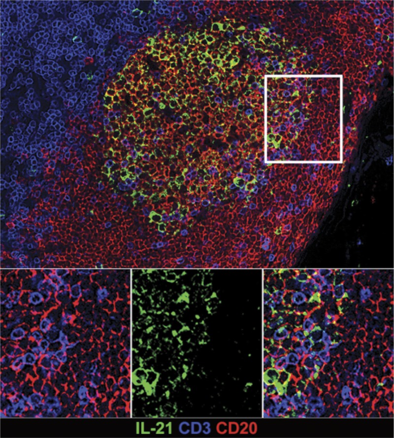
In situ IL-21 staining in the GC. IL-21 expression is increased in hyperplastic follicles during AMR. Representative immunofluorescence image of GC staining with CD3 (blue), CD20 (red), IL-21 (green) in lymph node of rhesus macaque.

In situ IL-21 staining in the GC. IL-21 expression is increased in hyperplastic follicles during AMR. Representative immunofluorescence image of GC staining with CD3 (blue), CD20 (red), IL-21 (green) in lymph node of rhesus macaque.