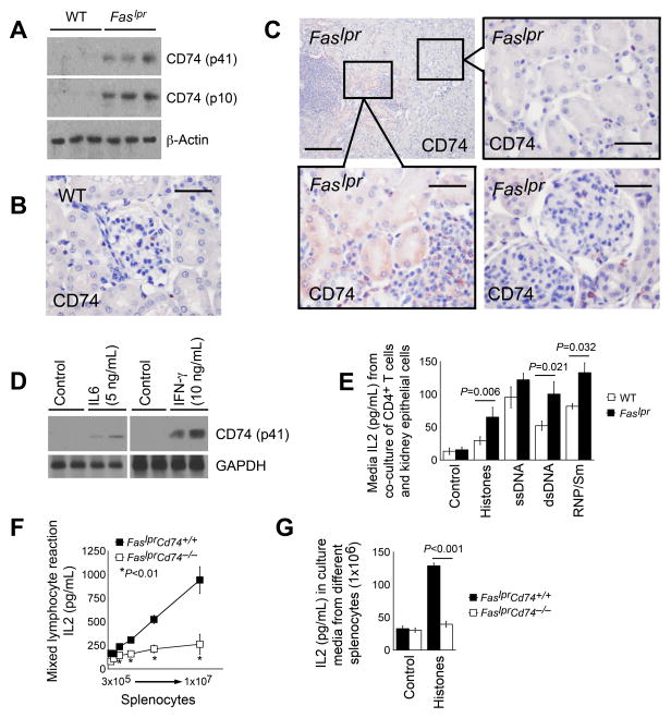
Figure 1.
CD74 expression and antigen presentation activity in Faslpr mice. A. Immunoblot detected CD74 expression, both the p41 full-length products and p10 processed fragments, in kidney extracts from 24-week-old WT and Faslpr mice. B. Immunostaining to detect CD74 (p41) expression in kidneys from 24-week-old WT mice. Scale: 50 μm. C. Immunostaining detected CD74 (p41) expression in kidneys from 24-week-old Faslpr mice. Scale: 200 μm and 50 μm, inset scale: 50 μm. D. CD74 (p41) expression in kidney TECs before and after stimulation with IL6 and IFN-γ. E. CD4+ T cell culture medium IL2 levels after activation with TECs from 24-week-old WT and Faslpr mice in the presence or absence of autoantigens as indicated. F. Mixed lymphocyte reactions of presenter splenocytes from 24-week-old FaslprCd74+/+ and FaslprCd74−/− mice and responder cells from bm12 mice. G. Autoantigen histone presentation assay. ELISA determined IL2 levels in splenocytes from 24-week-old FaslprCd74+/+ and FaslprCd74−/− mice treated with and without histone. Data in E–G are representative of three independent experiments.

