
FIGURE 1. OGG-1 responds to hyperoxic DNA damage and inflammation in lung cells.
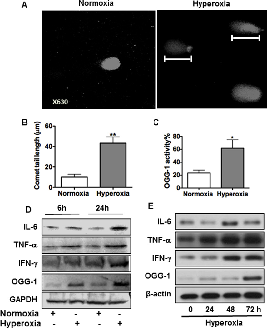
MLE-12 cells were incubated in room air or 95% O2. (A) DNA strand breaks were detected by a comet assay through measuring tail length (indicated by rulers) with confocal laser scanning fluorescence microscopy (CLSM). (B) Tail lengths were markedly increased in lung cells by hyperoxia compared to the control (P<0.001). (C) OGG-1 activity under 24 h hyperoxia was determined by incision enzymatic assay. (D) Increased inflammatory responses in MLE-12 cells after 6 h or 24 h exposure to hyperoxia by immunoblotting analysis. (E) Inflammatory responses in mice increased with exposure time (24, 48 and 72 h) by immunoblotting analysis. Data were representative of three experiments with similar results (student t-test, *p< 0.05, **p< 0.01).