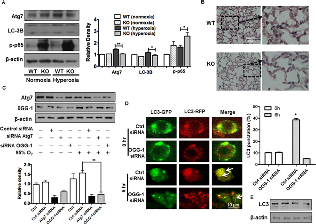
FIGURE 3. ogg-1 KO mice exhibit impaired autophagy under hyperoxia.

(A) Immunoblotting analysis of Atg7, p-NF-κB and LC3 in lungs of ogg-1 KO and WT mice (n=6) exposed to hyperoxia for 48 h. (B) Decreased expression of Atg7 in ogg-1 KO mice by immunohistochemistry. (C) Decreased Atg7 in MLE-12 cells after 48 h exposure to hyperoxia by immunoblotting analysis. Gel data were quantified using densitometry with Image J. (D) Tandem GFP-RFP-LC3 plasmids and OGG-1 siRNA were transfected to MH-S cells and then cells were exposed to hyperoxia for 6 h. Arrows indicate LC3 puncta. Data were representative of three experiments with similar results (student t-test, *p< 0.05, **p< 0.01). (E) Immunoblot analysis of LC3 with RFP-GFP-LC3/siOGG-1 transfected to MLE-12 cells that were exposed to hyperoxia for 6 h (lane 1: Ctrl siRNA 0 h; lane 2:OGG-1 siRNA 0 h; lane 3: Ctrl siRNA 6 h; lane 4: OGG-1 siRNA 6 h).
