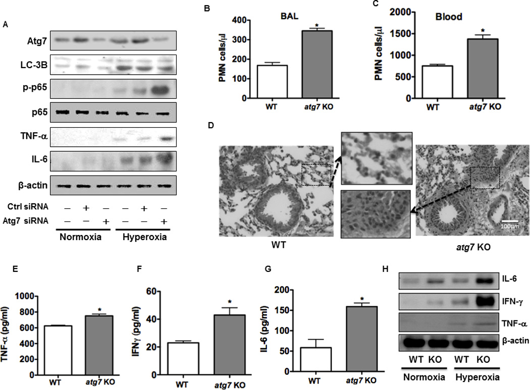
FIGURE 5. Atg7 deficiency contributes to intensified inflammatory responses under hyperoxia in vitro and in vivo.

(A) Increased expression of cytokines (IL-6, TNF-α) and p-NF-κB after knocking down Atg7 with siRNA in MLE-12 cells. (B) and (C) PMN infiltration and inflammatory response were increased in the lung (B) and blood (C) of atg7 KO mice compared to WT mice. (D) Increased lung injury and inflammation as assessed by H&E staining. (E)-(G) Increased inflammatory cytokines in BAL fluid of atg7 KO mice compared to WT mice by ELISA. (H) Increased expression of inflammatory cytokines in the lungs of atg7 KO mice (n=6) compared to WT mice after 48 h hyperoxic exposure by immunoblotting analysis. Data were representative of three experiments with similar results (student t-test, *p< 0.05).
