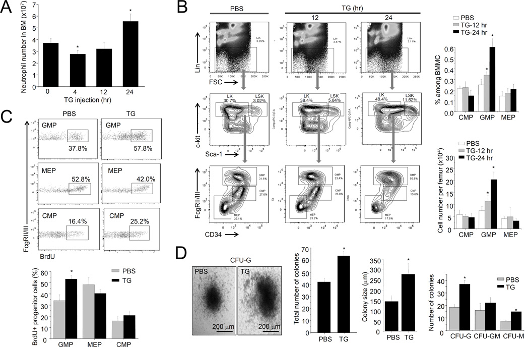
Figure 1. TG-induced sterile inflammation leads to increased progenitor cell proliferation in the BM.
(A) The number of neutrophils in the BM was measured using the Wright-Giemsa staining method. Data shown are means ± SD of n=5 mice. * p<0.01 versus control. (B) Flow cytometry-based lineage analysis of the BM cells. The percentage of each cell population among BM-derived mononuclear cells (BMMCs), and the absolute cell number per femur, are shown. Data shown are means ± SD of n=5 mice. * p<0.01 versus control (PBS treated mice). (C) Measurement of cycling cells in each progenitor population by incorporation of BrdU. Data shown are means ± SD of n=5 mice. * p<0.01 versus control. (D) The number of myeloid progenitors analyzed using an in vitro CFU-GM colony-forming assay. Data are means ± SD of n=5 mice. Representative pictures of cell clusters/colonies are shown.

