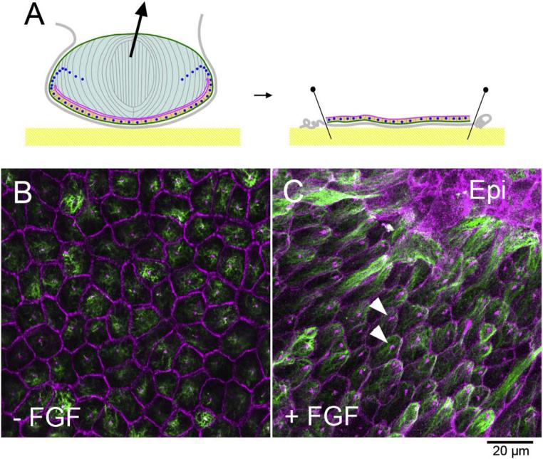
Fig. 2. Lens epithelial explants.
A Lens capsule with attached epithelial cells is pinned onto the bottom of culture dish. B, C Explant immunostained with β-catenin/pericentrin (purple, to show cell boundaries and centrosomes) and acetyl-tubulin (green, to detect cilia and microtubules) antibodies. Without FGF lens epithelial cells maintain cobblestone-like appearance (B). FGF induces groups of cells to elongate into fibers, whereas other groups of cells retain epithelial (epi) phenotype (C). The elongating fibers have hexagonal apical surfaces and cilia/centrosomes (arrowheads) are polarised towards the islands of epithelial cells (C).

