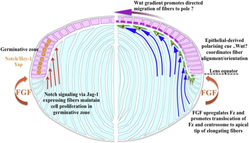
Fig. 3.
Proposed model for interactions between the epithelial-fiber (pink and light blue, respectively) compartments and key signaling pathways that regulate their dynamics. Right hand side of the diagram (Wnt-Frizzled signaling) shows that when epithelial cells shift below the lens equator, fiber differentiation is initiated in response to FGF in the posterior segment. This includes upregulation of Wnt-Fz signaling components and pathway activation that plays a key role in fiber elongation and differentiation (Dawes et al., 2013). This response also involves translocation of Fz (green) and the centrosome (red spot) to the apical tip of fibers as they elongate, and we propose this acts as the organizing center for cytoskeletal assembly and dynamics. Early in the elongation process the fibers have concave curvature (concave blue arrows) as they orient towards overlying epithelia in response to an epithelial-derived polarizing cue, possibly a Wnt (pink-purple dashed line in epithelium). As elongation progresses, the fibers take on convex curvature (convex blue arrows) as they undergo directed migration towards the pole, possibly in response to a Wnt concentration gradient. Left hand side shows that where the Jag-1 expressing cortical fibers (orange arrows) abut the epithelium determines the germinative zone, with nuclear localization of Notch-Jagged effector, Hey-1 and nuclear localization of Yap (orange star). Diagram modified from original in Dawes et al., 2014.

