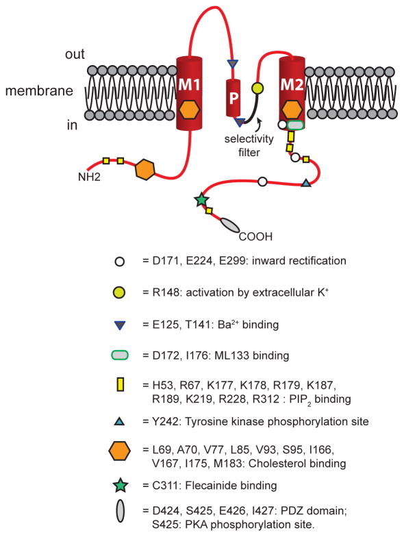
Figure 1.
Structure of KIR2 channels. Shown is a schematic representation of one KIR2 channel subunit positioned in the lipid bilayer of a cell membrane as shown. These channels have two membrane spanning helical domains, denoted M1 and M2 connected by a P-loop that contains a helical domain (P in the drawing). The channel’s pore is formed by the P-loop and M2, with the selectivity filter sequence highlighted in the drawing. Cytosolic amino (NH2) and carboxy (COOH) termini are also shown. Approximate locations of sites where regulatory molecules interact with the channel protein are shown as indicated. See text for references and more information.

