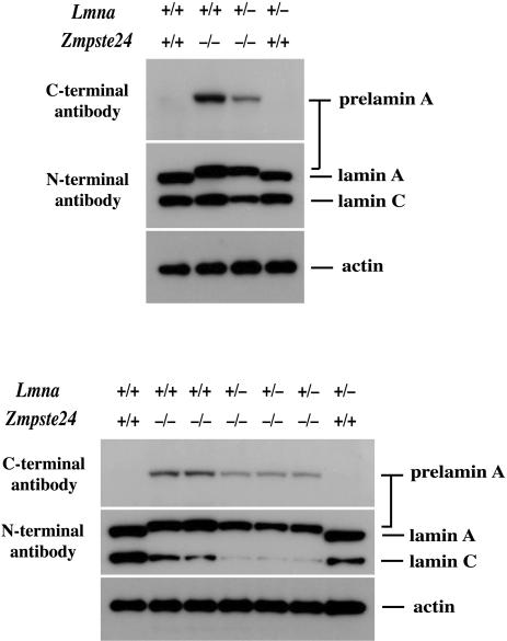
Fig. 4.
Western blots of extracts from wild-type, Zmpste24–/–, and Zmpste24–/–Lmna+/– MEFs with a carboxyl (C)-terminal prelamin A antibody and an amino (N)-terminal lamin A/C antibody. Analyses from two independent experiments, with cells prepared from two different sets of embryos, are shown. Protein loading was assessed with an antibody against β-actin. Densitometry showed a 58.4 ± 4.1% reduction in prelamin A and a 78 ± 4.9% decrease in lamin C in the Zmpste24–/–Lmna+/– cells (n = 4) relative to the Zmpste24–/– cells.

