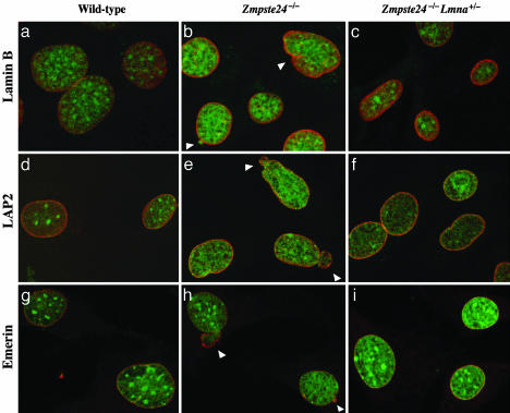
Fig. 5.
Analysis of nuclear shape in wild-type, Zmpste24–/–, and Zmpste24–/–Lmna+/– MEFs by laser-scanning fluorescence microscopy. The nuclear envelope was visualized with antibodies to lamin B (a–c), LAP2 (d–f), or emerin (g–i) (red), and DNA was visualized with SYTOX Green staining (green). White arrowheads indicate blebs.

