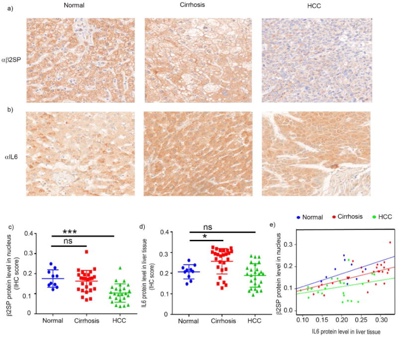
Fig. 1. Nuclear β2SP accumulation is inversely related to IL6 level in HCC tissues.

(a and b) Representative photomicrographs of β2SP (a) and IL6 (b) immunohistochemical staining in normal, cirrhotic, and HCC tissues. (c and d) Quantification of staining inetsity. Nuclear β2SP and IL6 protein levels were scored using inFORM software. (e) Robust linear regression using Huber's M estimator (Huber 1981) was applied to compare the correlation between IL6 with nuclear β2SP by cancer stage. Data in panels c–d are the means ± SEMs. *p < 0.05; **p < 0.01; ***p < 0.001; ****p < 0.0001, one-way ANOVA.
