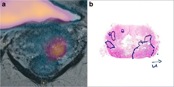
Fig. 3.

18F-Choline PET frame at 12.5 min fused with T2-weighted image (a). Focal tracer uptake is seen in the left lateral lobe. Histology confirmed tumor at this region with Gleason score 4 + 3 (b). No visible uptake was seen in the contralateral, lower grade tumor tissue (Gleason score 3 + 4)
