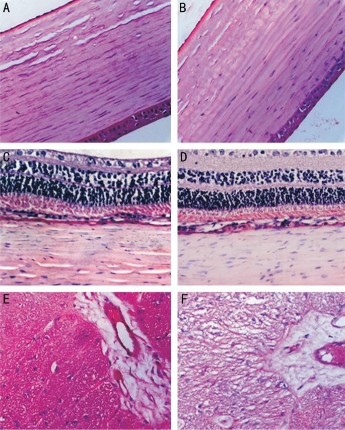
Figure 3. Photomicrographs of tissues in rabbit eye hematoxylin & eosin.

Panels (A, C, and E) are sections of untreated eyes, while panels (B, D, and F) are those of treated eyes. A, B: The cornea immediately adjacent to the scleral treatment quadrant with integrated endothelium and without stromal edema and loss of keratocytes; C, D: Intact retina, choroid, and sclera; E, F: The adjacent optic nerve without fiber swelling and rupture (400×).
