Abstract
Introduction
Maxillofacial region in children is particularly vulnerable to animal bite injuries. These injuries may range from insignificant scratches to life-threatening neck and facial injuries. Children are the common victims, particularly of dog bites.
Materials and methods
Three cases of animal bite injuries in children with their clinical presentation and their management are being presented along with review of literature. Surgical management included cleansing and primary closure of the wound. Rabies and tetanus prophylaxis were given.
Discussion
The most common site of injury was the face. For the facial injuries, the most frequently affected area was the middle third (55%), also called as the “central target area.” The small stature of children, the disproportionate size of the head relative to the body, their willingness to bring their faces close to the animal, and limited motor skills to provide defense are believed to account for this. The resulting soft-tissue injuries can vary in relation to their extent. Treatment involved initial surgical exploration, and secondary repair later depending on the severity of the injury.
Conclusion
Prompt assessment and treatment can prevent most bite wound complications. Early management of such injuries usually guarantees satisfactory outcome. Prevention strategies include close supervision of child-dog interactions, better reporting of bites, etc.
How to cite this article
Agrawal A, Kumar P, Singhal R, Singh V, Bhagol A. Animal Bite Injuries in Children: Review of Literature and Case Series. Int J Clin Pediatr Dent 2017;10(1):67-72.
Keywords: Animal bite injuries, Dog bites, Facial trauma.
INTRODUCTION
Facial trauma in children represents a significant medical and public health issue.1-4 A considerable proportion of skeletal and soft-tissue injuries of the face results from animal bite injuries, mostly due to dog bites.5 In the UK, it is estimated that dog attack injuries are responsible for an average of 250,000 minor injuries and emergency unit attendances each year,6 and in the USA, an average of 4.7 million dog bites occur each year7; many bites probably go unreported. Children, in particular, are more likely to experience dog bite injuries compared with adults, with children aged between 5 and 9 years considered to be the most at risk.6,8 Being the most exposed part of the body, the face is particularly vulnerable to such injuries.9-12 Among the victims of dog attacks, most studies showed a male preponderance.10,13,14 The types of wounds encountered range from insignificant scratches to life-threatening neck and facial injuries. The tissue defects may be superficial, but they can even cause amputations, including severe vascular and nerve or bony destruction. We present three cases of dog bite attacks in young children and their management.
CASE REPORTS
Case 1
A 3-year-old girl reported to the emergency department following an attack by a stray dog. She was otherwise fit and well, and had no relevant medical history or known allergies. A deep laceration wound was present extending from the left side of lower lip to the lower border of mandible (Fig. 1). A small laceration was present on the right nasolabial fold. Intraoral examination revealed that maxillary deciduous central incisors were slightly extruded and mobile. Her soft-tissue wounds were thoroughly debrided and irrigated with normal saline and hydrogen peroxide. The laceration was sutured with 4-0 round body vicryl and 4-0 reverse cutting prolene suture material (Fig. 2). The luxated maxillary incisors were stabilized with composite splinting. The parents were informed about the postoperative wound management. Tetanus and rabies prophylaxis were evaluated. The child was reviewed after 1 week and sutures were removed (Fig. 3). The patient was kept on regular follow-up for 3 months.
Fig. 1:
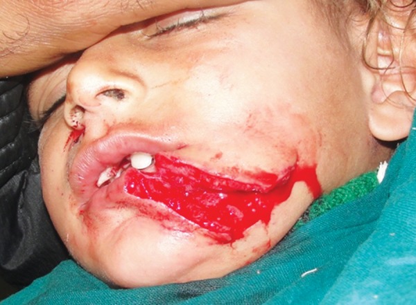
Deep lacerated wound in a 3-year-old girl
Fig. 2:
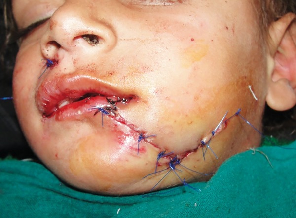
Sutured lacerated wound
Fig. 3:
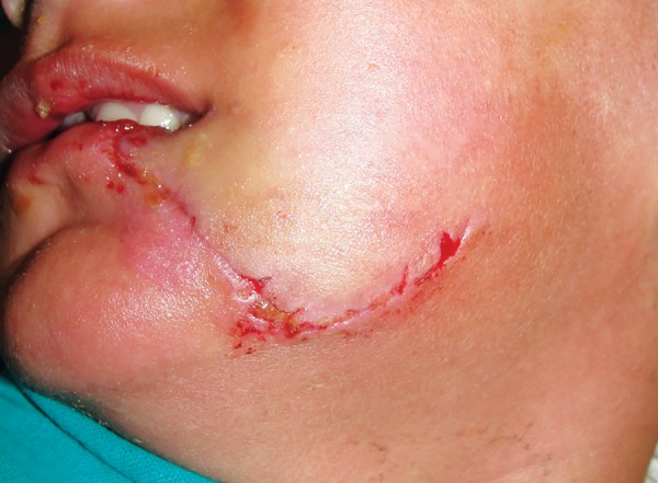
Follow-up picture after removal of sutures
Case 2
A 13-year-old boy reported to the emergency department following an attack by a stray dog. He was otherwise fit and well and had no relevant medical history or known allergies. A deep laceration wound was present on the left side of face extending 1 cm below and lateral to lower lip up to the lower border of mandible in the midline of face (Fig. 4). His soft-tissue wounds were thoroughly debrided and irrigated with normal saline and hydrogen peroxide. The laceration was sutured with vicryl and prolene suture material (Fig. 5). The parents were informed about the postoperative wound management. Tetanus and rabies prophylaxis were evaluated. The child was reviewed after 1 week and sutures were removed (Fig. 6). The patient was kept on regular follow-up for 3 months.
Fig. 4:
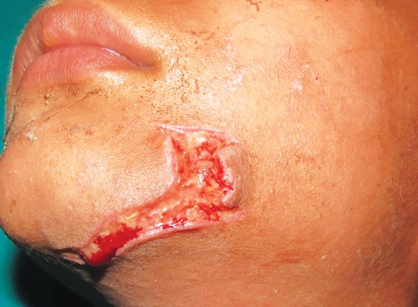
Lacerated wound in a 13-year-old boy
Fig. 5:
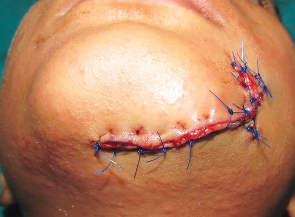
Sutured lacerated wound with vicryl and prolene
Fig. 6:
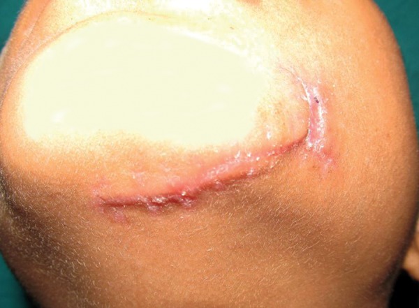
Follow-up picture showing healed wound
Case 3
A 6-year-old girl reported to the department with infected suture wound. Parents gave history of an attack by a stray dog, which was sutured by some private practitioner. The wound showed sign of infection with pus collection. She was otherwise fit and well and had no relevant medical history or known allergies. An infected laceration wound was present below the left eye extending up to the middle of the cheek. The sutures were removed and the margins of wound were refreshed with surgical blade (Fig. 7). Her soft-tissue wounds were thoroughly debrided and irrigated with normal saline and hydrogen peroxide. The laceration was sutured with vicryl and prolene suture material (Fig. 8). The parents were informed about the postoperative wound management. Tetanus and rabies prophylaxis were evaluated. The child was reviewed after 1 week and sutures were removed. The patient was kept on regular follow-up for 3 months.
Fig. 7:
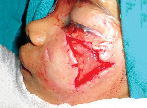
Lacerated wound in a 6-year-old girl
Fig. 8:
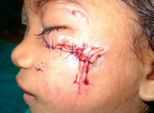
Sutured lacerated wound with vicryl and prolene material
DISCUSSION
Animal bites have been a major public health problem. Children are the most common victims, particularly of dog bites.15 The most common site of injury was the face.9-12 For the facial injuries, the most frequently affected area was the middle-third (55%).13 This reflects the findings of Palmer and Rees who called this the “central target area.”16 The small stature of children, the disproportionate size of the head relative to the body, their willingness to bring their faces close to the animal, and limited motor skills to provide defense are believed to account for this.4,17
A study showed that the risk factors for dog attacks include school-aged children (but highest rate of serious injury from dog bite is in children under 5 years of age),18 male, households with dogs, certain breeds (German shepherds, bull terriers, blue/red heelers, dobermans, and rottweilers), and male dogs. Most of the cases involve a known dog (friends, neighbors) and family pet.19
Dog bites are commonly associated with soft-tissue injury to the face, but rarely result in facial fractures.1,4,19,20 The injuries to the soft tissues are designated into three categories: Lacerations, punctures, and avulsions (tissue loss). The resulting soft-tissue injuries can vary considerably in relation to their extent and depth.20 The actual incidence of facial fractures relating to dog attacks is currently unknown. Schalamon et al.,1 Karlson,3 and Palmer and Rees16 documented no maxillofacial fractures in their review of facial dog bite injuries, and Tu et al20 suggested that facial fractures may occur in less than 5% of dog attack incidents.1,3,16,20 When a maxillofacial fracture is encountered, the most frequent bones to be fractured are the orbital, nasal, and maxillary bones, constituting 78% of the documented dog bite facial fractures.20,21 The mechanism of injury in cases of maxillofacial fracture is thought to be the consequence of the mandible (or involved bone) being physically held by the dogs jaws, which is capable of delivering immense force to the area of bone contacted by the dog’s teeth. In some breeds of dog, the force produced has been measured to be in the region of 31,790 kPa.6,22,23 The resultant force generated creates a crush-type injury and fracture of the alveolar bone. Young children are especially vulnerable to this type of crush injury, since the maxillofacial skeleton is not completely mineralized, is thinner, and, therefore, considerably weaker compared with during adulthood.20 Additional injuries due to animal bite included facial nerve damage, lacrimal duct damage requiring stenting and reconstruction, ptosis from levator transection, and blood loss requiring transfusion.19
The severity of the wounds was assessed by Lack-mann’s classification9:
I. Superficial injury without involvement of muscle.
II. Deep injury with involvement of muscle.
III. Deep injury with involvement of muscle and tissue defect.
IVa. Stage III in combination with vascular or nerve injury.
IVb. Stage III in combination with bony involvement or organ defect.
The optimal management of these wounds is controversial. The management of dog bite injuries has evolved over the years. In the past, accepted surgical practice involved delayed closure or healing by secondary intention. It was thought that because of the risk of infection, dog bite msinjuries should not be closed primarily.9,24 Pinsolle et al25 reviewed their series of dog bite injuries between 1979 and 1980. Treatment involved initial surgical exploration, followed by daily dressing with hydrogen peroxide and secondary repair 2 to 7 days later depending on the severity of the injury. More recently, there has been a move to more early and definitive treatment, with authors advocating early washout and debridement of wounds and primary closure.13,15,26-29 These changes have arisen from findings that the infection rate increased if treatment was delayed following injury,30 that debride-ment reduced the incidence of infection by as much as 30-fold,30 and that primary treatment produced the best cosmetic and functional results.9,10,26,30,31 Current opinion advocates early surgical treatment with irrigation of the wound, minimal debridement, and direct closure where possible.9,10,13,16,32,33 Postoperatively, attention to patient counseling, dressings, ointment, cleaning, and scar revision help assure an optimal outcome for the traumatized tissue. Avulsive injuries with significant tissue loss represent the most difficult cases for definitive management and are also those most likely to require hospitalization.34 For traumatic avulsion involving the lip vermilion and the perioral composite soft tissue, even with injuries including delicate anatomic landmarks, healing by secondary intention can be instituted as the initial treatment of choice in younger patients, often providing optimal results.35
Our regimen of primary closure after careful debridement of necrotic tissue has been the favored procedure in almost all recent publications.15,26-29 Wound cleansing is essential. We irrigated wounds with hydrogen peroxide and saline.15 Topical antibiotics and iodine solutions are no longer recommended.5 The use of water-based, rather than alcohol-based antiseptic solutions that cannot be used without local anesthesia solutions, is suggested by other authors.36
Wound infection is the most common complication following these injuries. Some authors estimate an infection rate of up to 30% following animal bite injuries to the extremities.37,38 Most infections caused by mammalian bites are polymicrobial, with mixed aerobic and anaerobic species. Bacteriology of infected dog and cat bite wounds includes Pasteurella multocida, Staphylococcus aureus, Viridans streptococci, Capnocytophaga canimorsus, and oral anaerobes.19 Presenting symptoms are usually wound site pain with cellulitis and purulent drainage.19 In addition to local wound infection, other complications may occur, including lymphangitis, local abscess, septic arthritis, tenosynovitis, and osteomyelitis. Rare complications include endocarditis, meningitis, brain abscess, and sepsis with disseminated intravascular coagulation, especially in immunocompromised individuals.19
Management of infection can be divided into cleansing of the wound, antibiotic prophylaxis, and antibiotic treat-ment.15 Antibiotic therapy is indicated for infected bite wounds and fresh wounds considered at-risk for infection, such as extremely large wounds, large hematoma, and cat bites, that appear to be more infected than dog bites (37.5 and 14.9% respectively) and immunocompromised patients.19 Antibiotic therapy (a combination of amoxicillin and clavulanic acid) and other combinations of extended-spectrum penicillins with beta-lactamase inhibitors offer the best in vitro coverage of the pathogenic flora.39 In patients with allergy to penicillins, monotherapy with azithromycin seems to be an effective alternative.39 Amoxycillin-clavulanic acid at a dose of 875 + 125 mg, twice a day, by mouth, for adults and 25 mg/ kg, twice a day, by mouth, for children seems to be the best regimen for prophylaxis in bite wound. Alternatively, azithromycin by mouth can be used (for adults 500 mg on day 1 and 250 mg a day for the next 4 days; for infants more than 6 months old, 10 mg/kg on day 1 followed by 5 mg/kg for the next 4 days).15 In case of slow recovery or no improvement, simultaneous lymphadenopathy, or pneumonia, S. aureus or Francisella tularensis should be suspected; ciprofloxacin is recommended.19 Prophylactic antibiotics are recommended for 5 to 7 days.15,40 Tetanus and rabies prophylaxis must be evaluated in all dog bites.
Metzger et al36 proposed the use of antibiotic prophylaxis for patients with comorbidities, high-risk injuries including cat bites, puncture wounds, bites older than 6 hours, extensive trauma to soft tissue, and bites in babies and infants. No antibiotic prophylaxis is necessary for scratch wounds or excoriations.14,41 Correira40 suggested the use of antibiotic prophylaxis also for patients with an edema at the site of the bite and for patients older than 50 years. Nearly all the patients in the study from Kountakis et al28 were given prophylactic antibiotics without regard to the severity of their injuries. Another study that focused on bacteriological background proposed antibiotic prophylaxis after bites by horses and birds.39
Prompt assessment and treatment can prevent most bite wound complications.19 Early management of such injuries usually guarantees satisfactory outcome. Prevention strategies include close supervision of child-dog interactions, public education about responsible dog ownership and dog bite prevention, stronger animal control laws, better resources for enforcement of these laws, and better reporting of bites.19 Anticipatory guidance by pediatric health care providers should attend to dog bite prevention. The need to improve community knowledge of rabies and the availability and affordability of rabies vaccine must be highlighted.19
Footnotes
Source of support: Nil
Conflict of interest: None
REFERENCES
- 1.Schalamon J, Ainoedhofer H, Singer G, Petnehazy T, Mayr J, Kiss K, Höllwarth ME. Analysis of dog bites in children who are younger than 17 years. Pediatrics. 2006 Mar;117(3):e374–e379. doi: 10.1542/peds.2005-1451. [DOI] [PubMed] [Google Scholar]
- 2.De Keuster T, Lamoureux J, Kahn A. Epidemiology of dog bites: a Belgian experience of canine behaviour and public health concerns. Vet J. 2006 Nov;172(3):482–487. doi: 10.1016/j.tvjl.2005.04.024. [DOI] [PubMed] [Google Scholar]
- 3.Karlson TA. The incidence of facial injuries from dog bites. . JAMA. 1984 Jun;251(24):3265–3267. [PubMed] [Google Scholar]
- 4.Shaikh ZS, Worrall SF. Epidemiology of facial trauma in a sample of patients aged 1-18 years. Injury. 2002 Oct;33(8):669–671. doi: 10.1016/s0020-1383(01)00201-7. [DOI] [PubMed] [Google Scholar]
- 5.Goldstein EJ. Bite wounds and infection. Clin Infect Dis. 1992 Mar;14(3):633–638. doi: 10.1093/clinids/14.3.633. [DOI] [PubMed] [Google Scholar]
- 6.Walker T, Modayil P, Cascarini L, Collyer JC. Dog bite fracture of the mandible in a 9 month old infant: a case report. Cases J. 2009 Jan;2(1):44. doi: 10.1186/1757-1626-2-44. [DOI] [PMC free article] [PubMed] [Google Scholar]
- 7.Sacks JJ, Kresnow M, Houston B. Dog bites: how big a problem? Inj Prev. 1996 Mar;2(1):52–54. doi: 10.1136/ip.2.1.52. [DOI] [PMC free article] [PubMed] [Google Scholar]
- 8.Weiss HB, Friedman DI, Coben JH. Incidence of dog bite injuries treated in emergency departments. JAMA. 1998 Jan;279(1):51–53. doi: 10.1001/jama.279.1.51. [DOI] [PubMed] [Google Scholar]
- 9.Lackmann G, Draf W, Isselstein G, Tollner U. Surgical treatment of facial dog bites injuries in children. J Craniomaxillofac Surg. 1992 Feb-Mar;20(2):81–86. doi: 10.1016/s1010-5182(05)80472-x. [DOI] [PubMed] [Google Scholar]
- 10.Mendez Gallart R, Gomez Tellado M, Somoza Argibay I, Liras Munoz J, Pais Pineiro E, Vela Nieto D. Mordeduras de perro. Analisis de 654 casos 10 anos. An Esp Pediatr. 2002 May;56(5):425–429. [PubMed] [Google Scholar]
- 11.Tuggle DW, Taylor DV, Stevens RJ. Dog bites in children. J Pediatr Surg. 1993 Jul;28(7):912–914. doi: 10.1016/0022-3468(93)90695-h. [DOI] [PubMed] [Google Scholar]
- 12.Wiseman NE, Chochinov H, Fraser V. Major dog attack injuries in children. J Pediatr Surg. 1983 Oct;18(5):533–536. doi: 10.1016/s0022-3468(83)80353-4. [DOI] [PubMed] [Google Scholar]
- 13.Akhtar N, Smith MJ, McKirdy S, Page RE. Surgical delay in the management of dog bite injuries in children, does it increase the risk of infection? J Plast Reconstr Aesthet Surg. 2006;59(1):80–85. doi: 10.1016/j.bjps.2005.05.005. [DOI] [PubMed] [Google Scholar]
- 14.Dire DJ. Cat bite wounds: risk factors for infection. Ann Emerg Med. 1991 Sep;20(9):973–979. doi: 10.1016/s0196-0644(05)82975-0. [DOI] [PubMed] [Google Scholar]
- 15.Kesting MR, Hölzle F, Pox C, Thurmuller P, Wolff KD. Animal bite injuries to the head: 132 cases. Br J Oral Maxillofac Surg. 2006 Jun;44(3):235–239. doi: 10.1016/j.bjoms.2005.06.015. [DOI] [PubMed] [Google Scholar]
- 16.Palmer J, Rees M. Dog bites of the face: a 15 year review. Br J Plast Surg. 1983 Jul;36(3):315–318. doi: 10.1016/s0007-1226(83)90051-6. [DOI] [PubMed] [Google Scholar]
- 17.Overall KL, Love M. Dog bites to human-demography, epidemiology, injury and risk. J Am Vet Med Assoc. 2001 Jun;218(12):1923–1934. doi: 10.2460/javma.2001.218.1923. [DOI] [PubMed] [Google Scholar]
- 18.Scheithauer MO, Rettinger G. Bite injuries in the head and neck area. HNO. 1997 Nov;45(11):891–897. doi: 10.1007/s001060050170. [DOI] [PubMed] [Google Scholar]
- 19.Abuabara A. A review of facial injuries due to dog bites. Med Oral Patol Oral Cir Bucal. 2006 Jul;11(4):E348–E350. [PubMed] [Google Scholar]
- 20.Tu AH, Girotto JA, Singh N, Dufresne CR, Robertson BC, Seyfer AE, Manson PN, Iliff N. Facial fractures from dog bite injuries. Plast Reconstr Surg. 2002 Apr;109(4):1259–1265. doi: 10.1097/00006534-200204010-00008. [DOI] [PubMed] [Google Scholar]
- 21.Brogan TV, Bratton SL, Dowd MD, Hegenbarth MA. Severe dog bites in children. Pediatrics. 1995 Nov;96(5 Pt 1):947–950. [PubMed] [Google Scholar]
- 22.Rosenthal DU. When K-9s cause chaos: an examination of police dog polices and their liabilities. NYL Sch J Hum Rts. 1993;11:279–310. [Google Scholar]
- 23.Presutti RJ. Prevention and treatment of dog bites. Am Fam Physician. 2001 Apr;63(8):1567–1572. [PubMed] [Google Scholar]
- 24.Jones RC, Shires GT. Bites and stings of animals and insects. Principles of surgery. New York: McGraw-Hill; 1979. pp. 232–242. [Google Scholar]
- 25.Pinsolle J, Phan E, Coustal B. Les morsures de chien au niveau de la face. Ann Chir Plast Esthet. 1993 Aug;38(4):452–456. [PubMed] [Google Scholar]
- 26.Wolff KD. Management of animal bite injuries of the face: experience with 94 patients. J Oral Maxillofac Surg. 1998 Jul;56(7):838–843. doi: 10.1016/s0278-2391(98)90009-x. [DOI] [PubMed] [Google Scholar]
- 27.Javaid M, Feldberg L, Gipson M. Primary repair of dog bites to the face: 40 cases. J R Soc Med. 1998 Aug;91(8):414–416. doi: 10.1177/014107689809100804. [DOI] [PMC free article] [PubMed] [Google Scholar]
- 28.Kountakis SE, Chamblee SA, Maillard AA, Stiernberg CM. Animal bites to the head and neck. Ear Nose Throat J. 1998 Mar;77(3):216–220. [PubMed] [Google Scholar]
- 29.Mitchell RB, Nanez G, Wagner JD, Kelly J. Dog bites of the scalp, face, and neck in children. Laryngoscope. 2003 Mar;113(3):492–495. doi: 10.1097/00005537-200303000-00018. [DOI] [PubMed] [Google Scholar]
- 30.Callham ML. Treatment of common dog bites: infection risk factors. JACEP. 1978 Mar;7(3):83–87. doi: 10.1016/s0361-1124(78)80063-x. [DOI] [PubMed] [Google Scholar]
- 31.Gonnering RS. Orbital and periorbital dog bites. Adv Ophthalmic Plast Reconstr Surg. 1987;7:71–80. [PubMed] [Google Scholar]
- 32.Goldstein EJ, Citron DM, Finegold SM. Dog bite wounds and infection: a prospective clinical study. Ann Emerg Med. 1980 Oct;9(10):508–512. doi: 10.1016/s0196-0644(80)80188-0. [DOI] [PubMed] [Google Scholar]
- 33.Mcheik JN, Vergnes P, Bondonny JM. Treatment of facial dog bite injuries in children: a retrospective study. J Pediatr Surg. 2000 Apr;35:580–583. doi: 10.1053/jpsu.2000.0350580. [DOI] [PubMed] [Google Scholar]
- 34.Stefanopoulos PK, Tarantzopoulou AD. Facial bite wounds: management update. Int J Oral Maxillofac Surg. 2005 Jul;34(5):464–472. doi: 10.1016/j.ijom.2005.04.001. [DOI] [PubMed] [Google Scholar]
- 35.Rhee ST, Colville C, Buchman SR. Conservative management of large avulsions of the lip and local landmarks. Pediatr Emerg Care. 2004 Jan;20(1):40–42. doi: 10.1097/01.pec.0000106243.72265.f6. [DOI] [PubMed] [Google Scholar]
- 36.Metzger R, Kanz KG, Lackner CK, Mutschler W. After cat bite antibiotics are obligatory. Acute management of bite injuries. MMW Fortschr Med. 2002 May;144(18):46–49. [PubMed] [Google Scholar]
- 37.Baker MD, Moore SE. Human bites in children: a six-year experience. Am J Dis Child. 1987 Dec;141(12):1285–1290. doi: 10.1001/archpedi.1987.04460120047032. [DOI] [PubMed] [Google Scholar]
- 38.Strady A, Rouger C, Vernet V, Combremont AG, Remy G, Deville J, Chippaux C. Animal bites. Epidemiology and infection risks. Presse Med. 1988 Nov;17(42):2229–2233. [PubMed] [Google Scholar]
- 39.Goldstein EJ. Current concepts on animal bites: bacteriology and therapy. Curr Clin Top Infect Dis. 1999;19:99–111. [PubMed] [Google Scholar]
- 40.Correira K. Managing dog, cat, and human bite wounds. JAAPA. 2003 Apr;16(4):28–37. [PubMed] [Google Scholar]
- 41.Dire DJ, Hogan DE, Walker JS. Prophylactic oral antibiotics for low risk dog bite wounds. Pediatr Emerg Care. 1992 Aug;8(4):194–99. doi: 10.1097/00006565-199208000-00005. [DOI] [PubMed] [Google Scholar]


