
Figure 1. Cryoinjury induces cell death, myocardial dysfunction, and decreased cardiomyocyte cell cycle activity in neonatal mice.
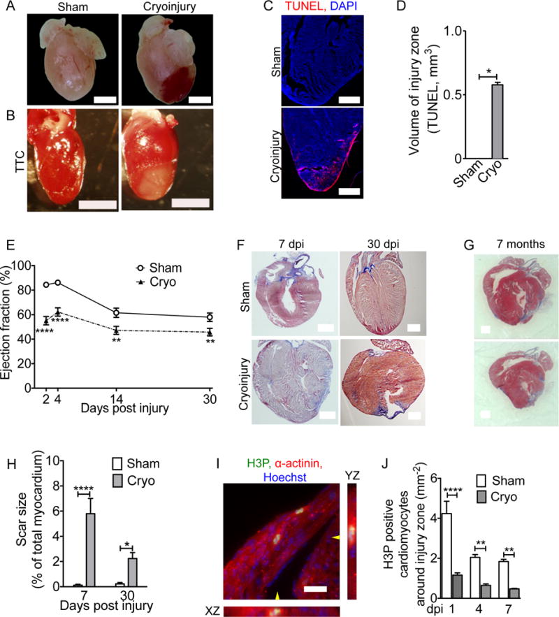
Mice underwent sham surgery or cryoinjury on day of life 1 (P1). (A) Hematoma at the injury site. (B) Vital staining with triphenyltetrazolium chloride (TTC) shows injury zone. (C,D) Myocardial cell death visualized by TUNEL staining. (E) Cryoinjury induces a sustained decrease in the ejection fraction. (F,G) AFOG-stained sections show that scar (blue) is formed within 7 dpi and present 30 days later (F). (G) Cyoinjury-induced scars, visualized on two sections of the same heart (500 μm apart) by Masson Trichrome staining, persist to 7 months after injury. (H) Quantification of scar size. (I) Two cardiomyocytes in M-phase visualized with antibodies against phosphorylated histone H3 (H3P) and α-actinin. The position of orthogonal reconstructions of the cardiomyocyte in the center are indicated with yellow arrowheads. (J) Quantification of M-phase cardiomyocytes in the region around the injury zone shows significant and sustained reduction after cryoinjury. Scale bars 1 mm (A,B,F,G), 20 μm (C,I). Statistical test by t-test (D) and ANOVA Bonferroni’s Multiple Comparison Test (E, H, J) * P < 0.05, ** P < 0.01, *** P < 0.001, **** P < 0.0001.