Figure 6.
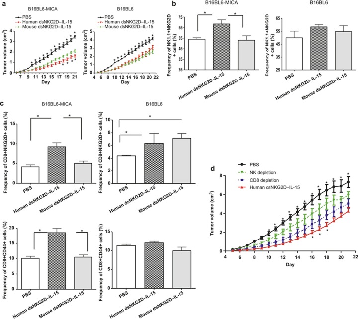
Anti-tumor effects of human dsNKG2D–IL-15depended mainly on NK cells. (a) B16BL6–MICA or B16 cells were injected into the C57BL/6 mice (n = 6, 2 × 10⁶ cells). Human dsNKG2D–IL-15 (60 μg), mouse dsNKG2D–IL-15 (60 μg), or PBS was also intraperitoneally injected daily into these mice from days 5 to 21. Tumor growth was measured daily and was shown as the means ± SD. The upper asterisks represent the comparison between human dsNKG2D–IL-15 and PBS, and the lower asterisks represent the comparison between human dsNKG2D–IL-15 and mouse dsNKG2D–IL-15. All mice were killed on day 22, and their spleens were collected. The frequencies of splenic NK1.1+NKG2D+ cells (b), CD8+NKG2D+ T cells and CD8+CD44+ T cells (c) were detected by flow cytometry. *P < 0.05, **P < 0.01. (d) Growth curve of B16-MICA transplanted tumors in mice treated with human dsNKG2D–IL-15 from day 5 following either NK or CD8+ T cell depletion (n = 5). The upper asterisks represent the comparison between human dsNKG2D–IL-15 and NK cell depletion, and the lower asterisks represent the comparison between human dsNKG2D–IL-15 and CD8+ T-cell depletion. The experiments were performed twice.

