
Figure 2.
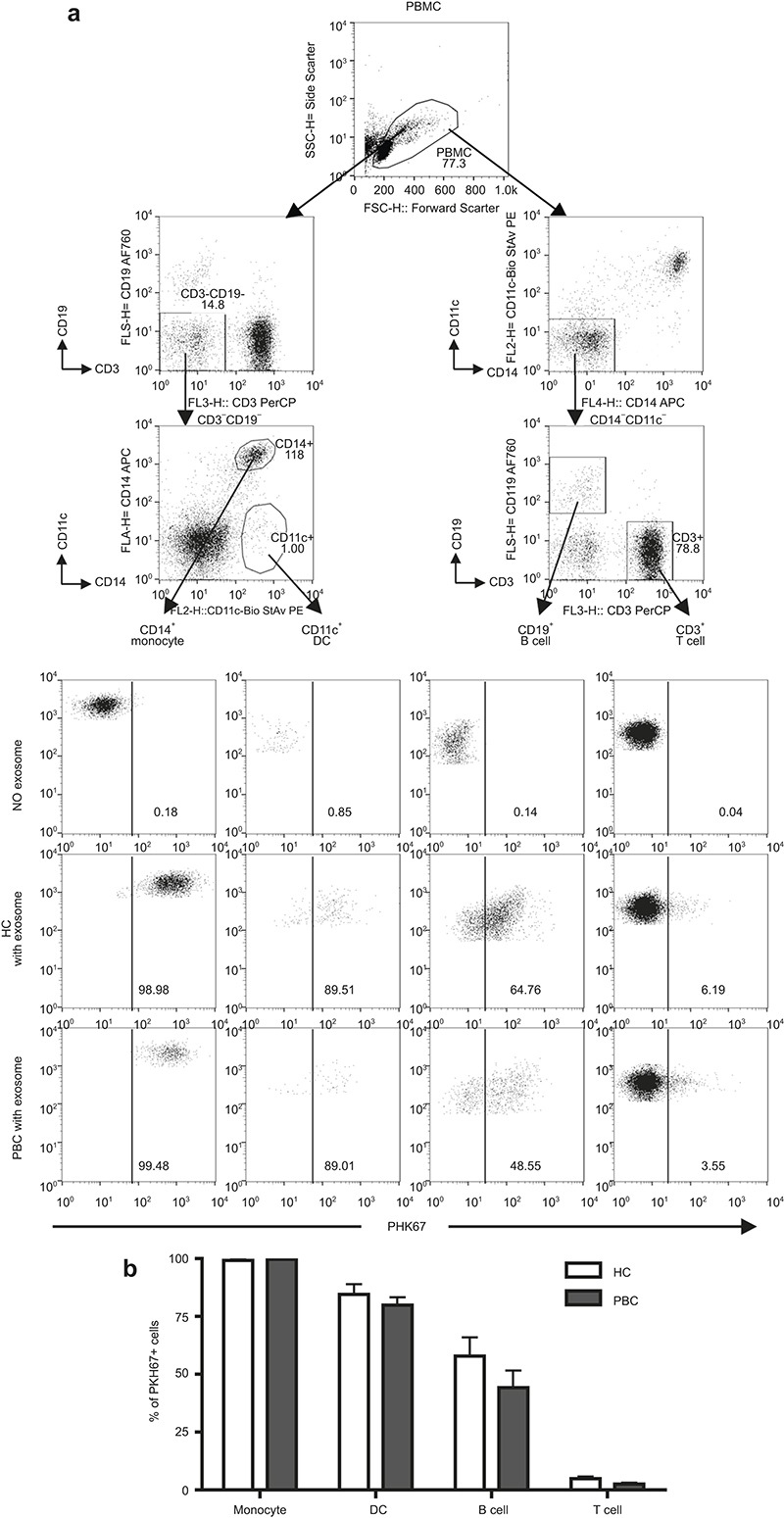
Uptake of exosomes by PBMCs. Exosomes were labeled with PKH-67 dye and cultured with freshly isolated 1 × 10⁶ PBMCs for 16 h. (a) A representative pattern of PKH-67+ cells in each subpopulation was analyzed by flow cytometry. (b) Frequency of PKH-67+ cells in CD14+ monocytes, CD11c+ DCs, CD19+ B cells, and CD3+ T cells. Data are shown as mean ± SEM.