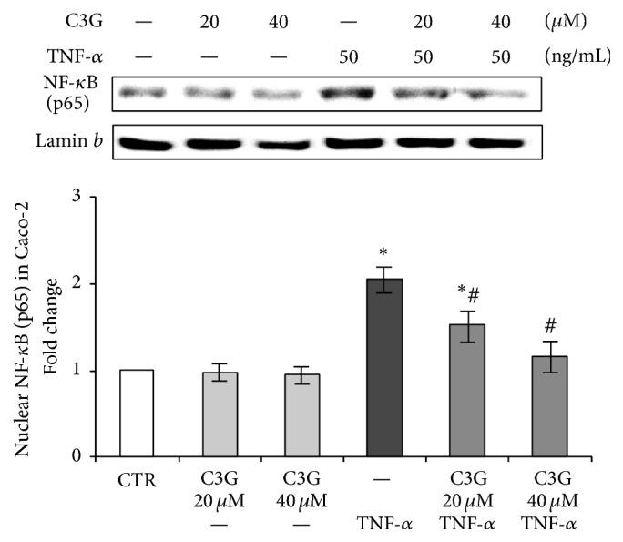
Figure 2.

Nuclear NF-κB (p65) in Caco-2 cells. The Caco-2 monolayer was pretreated for 24 hours with C3G (20 or 40 μM) and subsequently exposed to 50 ng/mL TNF-α for 1 hour. Cultures treated with the vehicle alone (0.1% DMSO) were used as controls (CTR). Caco-2 cell nuclear lysates were analyzed by western blot, and nuclear localization of the p65 protein was evaluated. Results are reported as fold change against control and expressed as mean ± SD of three independent experiments. NF-κB (p65) intensity values were normalized to the corresponding Lamin b value. ∗p < 0.05 versus CTR; #p < 0.05 versus TNF-α.
