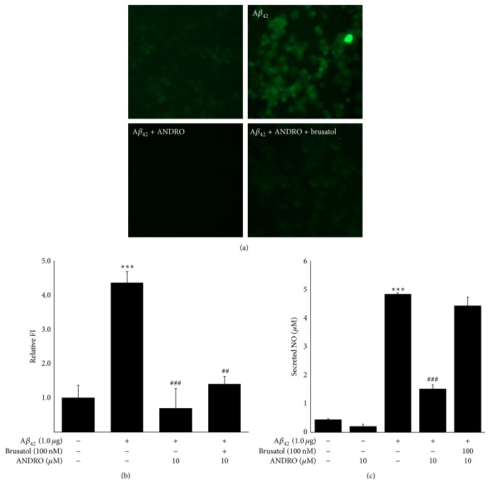
Figure 4.
Effect of andrographolide and/or brusatol on Aβ42 expression in BV-2 cells. BV-2 cells were transfected with Aβ42 plasmids for 12 h. The medium was discarded and replaced with phenol red-free medium in the absence or presence of 100 nM brusatol. After 2 h, the medium was replaced with fresh medium containing 1, 5, or 10 μM of andrographolide. The abbreviation of andrographolide, ANDRO, was used. (a) The microglial clearance of Aβ42 was visualized by fluorescence microscope imaging. (b) The relative fluorescence intensity of Aβ42 was quantified by Image J software. The data are presented as the means ± SE of triplicates. The significance presented as ∗∗∗p < 0.001 compared with control group and ##p < 0.01 and ###p < 0.001 compared with the Aβ42-transfected group. (c) The secreted NO levels in the medium of BV-2 cells were measured using the Griess method. The data are presented as the means ± SE of triplicates. The significance presented as ∗∗∗p < 0.001 compared with control group and ###p < 0.001 compared with the Aβ42-transfected group.

