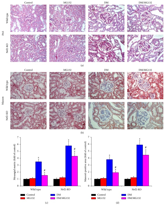
Figure 2.
Effects of MG132 on diabetes-induced morphological changes were examined with PAS (a) and Masson's trichrome staining (b, ×400) in all mice. Mesangial matrix expansion (c) was quantified from PAS staining and fibrosis accumulation (d) was quantified from Masson's trichrome staining. Data are presented as mean ± SD. ∗p < 0.05 versus WT/control or Nrf2-KO/control correspondingly; #p < 0.05 versus WT/DM or Nrf2-KO/DM correspondingly.

