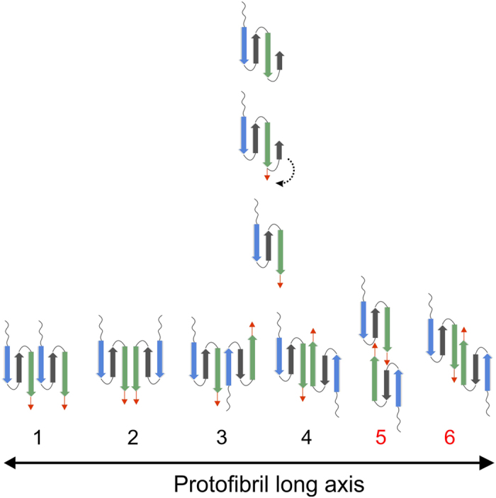
Figure 5. Schematic representation of possible medin dimers.

Cartoon illustration of proposed C-terminal strand detachment and subsequent dimer formation through interaction of the amyloidogenic first and third strands (blue and green respectively) as defined by Aggrescan3D (see Fig. 4e). Options 1–4 could involve assembly through pre-formed strands; blue strand (N7-A13) and green strand (K30-I36) with optional involvement of the C-terminal residues 36–50 (red arrow), whereas options 5 and 6 requires stabilisation by the ‘zipping back’ of the C-terminus to form a longer strand encompassing residues 30–50 (green strand and red arrow).
