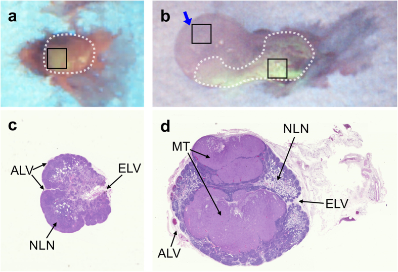
Figure 4.
Post-operative analysis: ex vivo FL validation of the resected SLNs from (a) control and (b) tumor-bearing rabbits. The blue arrow in (b) indicates the direction of FL imaging available during in vivo surgical guidance. The black rectangles indicate the regions-of-interest for FL intensity measurements. H&E stained sections are shown for the corresponding (c) control and (d) tumor-bearing SLN: MT-metastatic tumor, EL-efferent lymph vessel, ALV-afferent lymph vessel, NLN-normal lymph node tissue.

