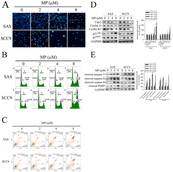
Figure 2. MP induces apoptosis and G2/M phase cell cycle arrest in SAS and SCC9 cells.
Cells were treated with different concentration of MP (0, 2, 4 and 8 μM) for 24 h. (A) Morphological changes in cells including nuclei condensation and fragmentation were observed by DAPI staining under a fluorescence microscope. (B) Cell cycle was analysed by PI staining and flow cytometry. Sub-G1, G0/G1, S and G2/M indicate different cell cycle phases. (C) Apoptotic cells were stained with Annexin V-FITC/PI and analyzed by flow cytometry. (D) The left panel shows representative Western blots for the effect of MP on expression of cell cycle regulatory proteins (Cdc2, Cyclin A, Cyclin B1, p21 Cip1, and p27 Kip1). Bar graphs represent the relative density of each band normalized to GAPDH (right panel). (E) The left panel shows representative Western blots for the effect of MP on expression of apoptosis-related proteins (cleaved caspase-3, -8, -9 and cleaved PARP). Bar graphs represent the relative density of each band normalized to GAPDH (right panel). Values represent the mean ± SD of 3 independent experiments. *P < 0.05, compared with the control (0 μM).

