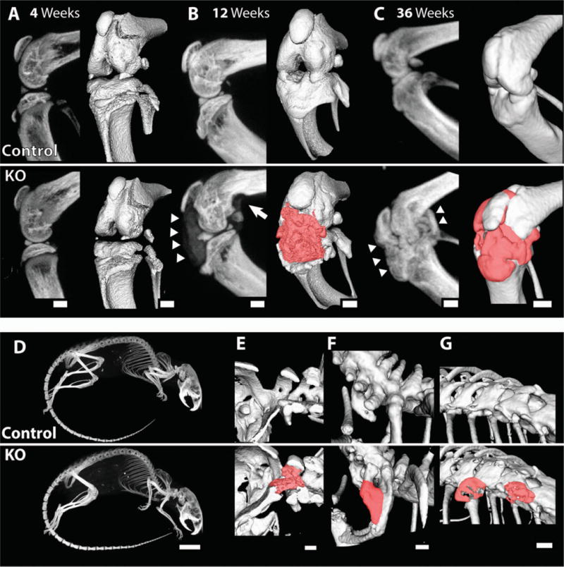
Figure 4.

Calcified nodules in the knee and spine of 12-week-old and 36-week-old cartilage-specific mitogen-specific gene 6–knockout (KO) mice and control mice. Control and KO mice were scanned by micro–computed tomography at the indicated ages. A–C, Sagittal plane maximum-intensity projection (MIP) images (left) and 3-dimensional isosurface images (right) obtained at age 4 weeks (A), 12 weeks (B), and 36 weeks (C), showing ectopic calcified tissue (arrowheads and red manual contrast versus white for bone) and bone erosion (arrow) in the KO mice. D, MIP images of 36-week-old mice. E–G, Three-dimensional isosurface images, showing the presence of calcified material (red manual contrast versus white for bone) in some of the 36-week-old KO animals at the base of the skull (E), as a fusion between the C7 and T1 vertebrae (F), and between the T10 and T11 vertebrae (G). Images are representative of 3 or more mice per group. Bars = 1 mm in A–C and E–G; 1 cm in D.
